Three-dimensional structure of the DNA-binding domain of the fructose repressor from Escherichia coli by 1H and 15N NMR.
Penin, F., Geourjon, C., Montserret, R., Bockmann, A., Lesage, A., Yang, Y.S., Bonod-Bidaud, C., Cortay, J.C., Negre, D., Cozzone, A.J., Deleage, G.(1997) J Mol Biol 270: 496-510
- PubMed: 9237914
- DOI: https://doi.org/10.1006/jmbi.1997.1123
- Primary Citation of Related Structures:
1UXC, 1UXD - PubMed Abstract:
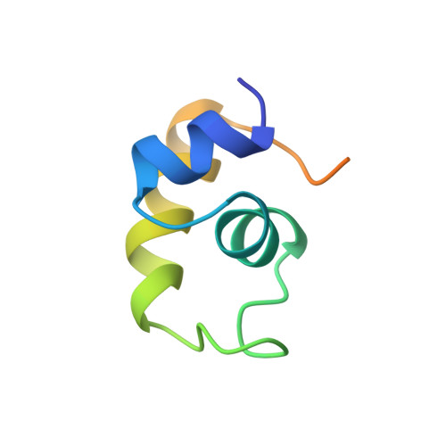
FruR is an Escherichia coli transcriptional regulator that belongs to the LacI DNA-binding protein family. By using 1H and 15N NMR spectroscopy, we have determined the three-dimensional solution structure of the FruR N-terminal DNA-binding domain consisting of 57 amino acid residues. A total of 809 NMR-derived distances and 54 dihedral angle constraints have been used for molecular modelling with the X-PLOR program. The resulting set of calculated structures presents an average root-mean-square deviation of 0.37 A at the main-chain level for the first 47 residues. This highly defined N-terminal part of the structure reveals a similar topology for the three alpha-helices when compared to the 3D structures of LacI and PurR counterparts. The most striking difference lies in the connection between helix II and helix III, in which three additional residues are present in FruR. This connecting segment is well structured and contains a type III turn. Apart from hydrophobic interactions of non-polar residues with the core of the domain, this connecting segment is stabilised by several hydrogen bonds and by the aromatic ring stacking between Tyr19 of helix II and Tyr28 of the turn. The region containing the putative "hinge helix" (helix IV), that has been described in PurR-DNA complex to make specific base contacts in the minor groove of DNA, is unfolded. Examination of hydrogen bonds highlights the importance of homologous residues that seem to be conserved for their ability to fulfill helix N and C-capping roles in the LacI repressor family.
Organizational Affiliation:
Institut de Biologie et Chimie des Proteines, Centre National de la Recherche Scientifique, Lyon, France.