1UX6

Structure of a thrombospondin C-terminal fragment reveals a novel calcium core in the type 3 repeats


Experimental Data Snapshot

  • Method: X-RAY DIFFRACTION
  • Resolution: 1.90 Å
  • R-Value Free: 0.227 
  • R-Value Work: 0.199 
  • R-Value Observed: 0.199 

wwPDB Validation   3D Report Full Report


This is version 1.3 of the entry. See complete history


Literature

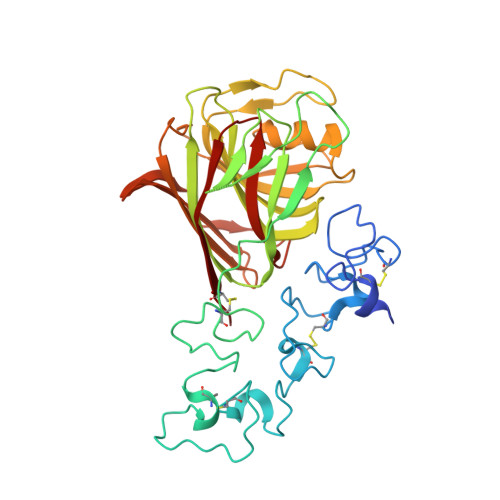
Structure of a Thrombospondin C-Terminal Fragment Reveals a Novel Calcium Core in the Type 3 Repeats

Kvansakul, M.Adams, J.C.Hohenester, E.

(2004) EMBO J 23: 1223

  • DOI: https://doi.org/10.1038/sj.emboj.7600166
  • Primary Citation of Related Structures:  
    1UX6

  • PubMed Abstract: 

    Thrombospondins (TSPs) are extracellular regulators of cell-matrix interactions and cell phenotype. The most highly conserved region of all TSPs are the calcium-binding type 3 (T3) repeats and the C-terminal globular domain (CTD). The crystal structure of a cell-binding TSP-1 fragment, spanning three T3 repeats and the CTD, reveals a compact assembly. The T3 repeats lack secondary structure and are organised around a core of calcium ions; two DxDxDGxxDxxD motifs per repeat each encapsulate two calcium ions in a novel arrangement. The CTD forms a lectin-like beta-sandwich and contains four strictly conserved calcium-binding sites. Disruption of the hairpin structure of T3 repeats 6 and 7 decreases protein secretion and stability. The availability for cell attachment of an RGD motif in T3 repeat 7 is modulated by calcium loading. The central architectural role of calcium explains how it is critical for the functions of the TSP C-terminal region. Mutations in the T3 repeats of TSP-5/COMP, which cause two human skeletal disorders, are predicted to disrupt the tertiary structure of the T3-CTD assembly.


  • Organizational Affiliation

    Department of Biological Sciences, Imperial College London, South Kensington Campus, London, UK.


Macromolecules
Find similar proteins by:  (by identity cutoff)  |  3D Structure
Entity ID: 1
MoleculeChains Sequence LengthOrganismDetailsImage
THROMBOSPONDIN-1350Homo sapiensMutation(s): 2 
UniProt & NIH Common Fund Data Resources
Find proteins for P07996 (Homo sapiens)
Explore P07996 
Go to UniProtKB:  P07996
PHAROS:  P07996
GTEx:  ENSG00000137801 
Entity Groups  
Sequence Clusters30% Identity50% Identity70% Identity90% Identity95% Identity100% Identity
UniProt GroupP07996
Sequence Annotations
Expand
  • Reference Sequence
Small Molecules
Ligands 1 Unique
IDChains Name / Formula / InChI Key2D Diagram3D Interactions
CA
Query on CA

Download Ideal Coordinates CCD File 
B [auth A]
C [auth A]
D [auth A]
E [auth A]
F [auth A]
B [auth A],
C [auth A],
D [auth A],
E [auth A],
F [auth A],
G [auth A],
H [auth A],
I [auth A],
J [auth A],
K [auth A],
L [auth A],
M [auth A],
N [auth A],
O [auth A],
P [auth A],
Q [auth A]
CALCIUM ION
Ca
BHPQYMZQTOCNFJ-UHFFFAOYSA-N
Experimental Data & Validation

Experimental Data

  • Method: X-RAY DIFFRACTION
  • Resolution: 1.90 Å
  • R-Value Free: 0.227 
  • R-Value Work: 0.199 
  • R-Value Observed: 0.199 
  • Space Group: H 3 2
Unit Cell:
Length ( Å )Angle ( ˚ )
a = 162.34α = 90
b = 162.34β = 90
c = 85.14γ = 120
Software Package:
Software NamePurpose
CNSrefinement
MOSFLMdata reduction
SCALAdata scaling
MLPHAREphasing

Structure Validation

View Full Validation Report


Entry History 

Deposition Data

Revision History  (Full details and data files)

  • Version 1.0: 2004-03-18
    Type: Initial release
  • Version 1.1: 2011-05-07
    Changes: Version format compliance
  • Version 1.2: 2011-07-13
    Changes: Version format compliance
  • Version 1.3: 2017-07-12
    Changes: Derived calculations