1UX5

Crystal Structures of a Formin Homology-2 domain reveal a flexibly tethered dimer architecture


Experimental Data Snapshot

  • Method: X-RAY DIFFRACTION
  • Resolution: 2.50 Å
  • R-Value Free: 0.251 
  • R-Value Work: 0.207 
  • R-Value Observed: 0.207 

wwPDB Validation   3D Report Full Report


This is version 1.3 of the entry. See complete history


Literature

Crystal Structures of a Formin Homology-2 Domain Reveal a Tethered Dimer Architecture

Xu, Y.Moseley, J.B.Sagot, I.Poy, F.Pellman, D.Goode, B.L.Eck, M.J.

(2004) Cell 116: 711

  • DOI: https://doi.org/10.1016/s0092-8674(04)00210-7
  • Primary Citation of Related Structures:  
    1UX4, 1UX5

  • PubMed Abstract: 

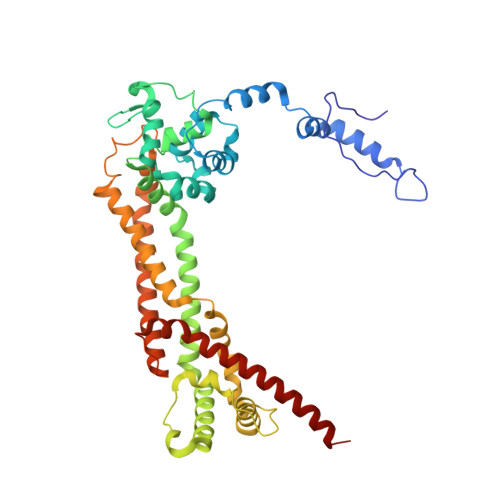
    Formin proteins participate in a wide range of cytoskeletal processes in all eukaryotes. The defining feature of formins is a highly conserved approximately 400 residue region, the Formin Homology-2 (FH2) domain, which has recently been found to nucleate actin filaments. Here we report crystal structures of the S. cerevesiae Bni1p FH2 domain. The mostly alpha-helical FH2 domain forms a unique "tethered dimer" in which two elongated actin binding heads are tied together at either end by an unusual lasso and linker structure. Biochemical and crystallographic observations indicate that the dimer is stable but flexible, with flexibility between the two halves of the dimer conferred by the linker segments. Although each half of the dimer is competent to interact with filament ends, the intact dimer is required for actin nucleation and processive capping. The tethered dimer architecture may allow formins to stair-step on the barbed end of an elongating nascent filament.


  • Organizational Affiliation

    Department of Cancer Biology, Dana-Farber Cancer Institute, Boston, MA 02115, USA.


Macromolecules
Find similar proteins by:  (by identity cutoff)  |  3D Structure
Entity ID: 1
MoleculeChains Sequence LengthOrganismDetailsImage
BNI1 PROTEIN411Saccharomyces cerevisiaeMutation(s): 0 
UniProt
Find proteins for P41832 (Saccharomyces cerevisiae (strain ATCC 204508 / S288c))
Explore P41832 
Go to UniProtKB:  P41832
Entity Groups  
Sequence Clusters30% Identity50% Identity70% Identity90% Identity95% Identity100% Identity
UniProt GroupP41832
Sequence Annotations
Expand
  • Reference Sequence
Experimental Data & Validation

Experimental Data

  • Method: X-RAY DIFFRACTION
  • Resolution: 2.50 Å
  • R-Value Free: 0.251 
  • R-Value Work: 0.207 
  • R-Value Observed: 0.207 
  • Space Group: P 61 2 2
Unit Cell:
Length ( Å )Angle ( ˚ )
a = 101.4α = 90
b = 101.4β = 90
c = 265.7γ = 120
Software Package:
Software NamePurpose
CNSrefinement
SCALEPACKdata scaling

Structure Validation

View Full Validation Report


Entry History 

Deposition Data

Revision History  (Full details and data files)

  • Version 1.0: 2004-03-11
    Type: Initial release
  • Version 1.1: 2011-05-07
    Changes: Version format compliance
  • Version 1.2: 2011-07-13
    Changes: Version format compliance
  • Version 1.3: 2019-05-08
    Changes: Advisory, Data collection, Experimental preparation, Other