Reassessment of Protein Stability, DNA Binding, and Protection of Mycobacterium Smegmatis Dps.
Ceci, P., Ilari, A., Falvo, E., Giangiacomo, L., Chiancone, E.(2005) J Biol Chem 280: 34776
- PubMed: 16030020
- DOI: https://doi.org/10.1074/jbc.M502343200
- Primary Citation of Related Structures:
1UVH - PubMed Abstract:
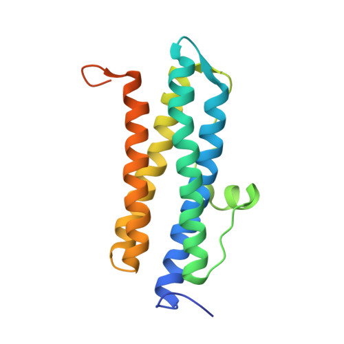
The structure and function of Mycobacterium smegmatis Dps (DNA-binding proteins from starved cells) and of the protein studied by Gupta and Chatterji, in which the C terminus that is used for binding DNA contains a histidine tag, have been characterized in parallel. The native dodecamer dissociated reversibly into dimers above pH 7.5 and below pH 6.0, with apparent pK(a) values of approximately 7.65 and 4.75; at pH approximately 4.0, dimers formed monomers. Based on structural analysis, the two dissociation steps have been attributed to breakage of the salt bridges between Glu(157) and Arg(99) located at the 3-fold symmetry axes and to protonation of Asp(66) hydrogen-bonded to Lys(36) across the dimer interface, respectively. The C-terminal tag did not affect subunit dissociation, but altered DNA binding dramatically. At neutral pH, protonation of the histidine tag promoted DNA condensation, whereas in the native C terminus, compensation of negative and positive charges led to DNA binding without condensation. This different mode of interaction with DNA has important functional consequences as indicated by the failure of the native protein to protect DNA from DNase-mediated cleavage and by the efficiency of the tagged protein in doing so as a result of DNA sequestration in the condensates. Chemical protection of DNA from oxidative damage is realized by Dps proteins in a multistep iron oxidation/uptake/mineralization process. Dimers have a decreased protection efficiency due to disruption of the dodecamer internal cavity, where iron is deposited and mineralized after oxidation at the ferroxidase center.
Organizational Affiliation:
National Research Council Institute of Molecular Biology and Pathology, Department of Biochemical Sciences "A. Rossi-Fanelli," University of Rome "La Sapienza," 00185 Rome, Italy.