1UTI

Mona/Gads SH3C in complex with HPK derived peptide


Experimental Data Snapshot

  • Method: X-RAY DIFFRACTION
  • Resolution: 1.50 Å
  • R-Value Free: 0.221 
  • R-Value Work: 0.207 
  • R-Value Observed: 0.208 

wwPDB Validation   3D Report Full Report


This is version 1.3 of the entry. See complete history


Literature

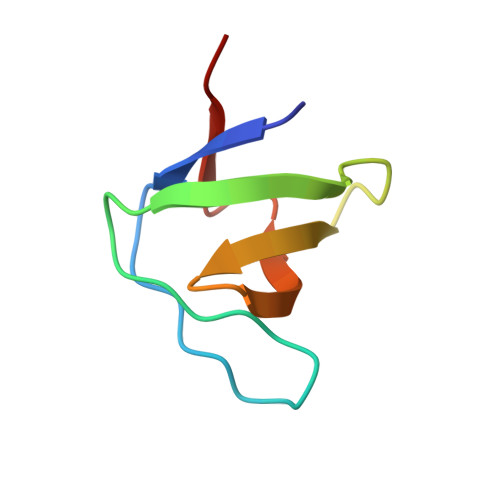
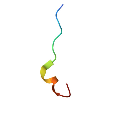
Mona/Gads Sh3C Binding to Hematopoietic Progenitor Kinase 1 (Hpk1) Combines an Atypical SH3 Binding Motif, R/Kxxk, with a Classical Pxxp Motif Embedded in a Polyproline Type II (Ppii) Helix

Lewitzky, M.Harkiolaki, M.Domart, M.C.Jones, E.Feller, S.M.

(2004) J Biol Chem 279: 28724

  • DOI: https://doi.org/10.1074/jbc.M402745200
  • Primary Citation of Related Structures:  
    1UTI

  • PubMed Abstract: 

    Hematopoietic progenitor kinase 1 (HPK1) is implicated in signaling downstream of the T cell receptor. Its non-catalytic, C-terminal half contains several prolinerich motifs, which have been shown to interact with different SH3 domain-containing adaptor proteins in vitro. One of these, Mona/Gads, was also shown to bind HPK1 in mouse T cells in vivo. The region of HPK1 that binds to the Mona/Gads C-terminal SH3 domain has been mapped and shows only very limited similarity to a recently identified high affinity binding motif in SLP-76, another T-cell adaptor. Using isothermal titration calorimetry and x-ray crystallography, the binding of the HPK1 motif to Mona/Gads SH3C has now been characterized in molecular detail. The results indicate that although charge interactions through an RXXK motif are essential for complex formation, a PXXP motif in HPK1 strongly complements binding. This unexpected binding mode therefore differs considerably from the previously described interaction of Mona/Gads SH3C with SLP-76. The crystal structure of the complex highlights the great versatility of SH3 domains, which allows interactions with very different proteins. This currently limits our ability to categorize SH3 binding properties by simple rules.


  • Organizational Affiliation

    Cancer Research UK Cell Signalling Group, Molecular Oncology Laboratory, Weatherall Institute of Molecular Medicine, University of Oxford, Oxford OX3 9DS, United Kingdom.


Macromolecules
Find similar proteins by:  (by identity cutoff)  |  3D Structure
Entity ID: 1
MoleculeChains Sequence LengthOrganismDetailsImage
GRB2-RELATED ADAPTOR PROTEIN 258Mus musculusMutation(s): 0 
UniProt
Find proteins for O89100 (Mus musculus)
Explore O89100 
Go to UniProtKB:  O89100
Entity Groups  
Sequence Clusters30% Identity50% Identity70% Identity90% Identity95% Identity100% Identity
UniProt GroupO89100
Sequence Annotations
Expand
  • Reference Sequence

Find similar proteins by:  Sequence   |   3D Structure  

Entity ID: 2
MoleculeChains Sequence LengthOrganismDetailsImage
MITOGEN-ACTIVATED PROTEIN KINASE KINASE KINASE KINASE 1B [auth D]16Mus musculusMutation(s): 0 
EC: 2.7.1.37
UniProt
Find proteins for P70218 (Mus musculus)
Explore P70218 
Go to UniProtKB:  P70218
Entity Groups  
Sequence Clusters30% Identity50% Identity70% Identity90% Identity95% Identity100% Identity
UniProt GroupP70218
Sequence Annotations
Expand
  • Reference Sequence
Experimental Data & Validation

Experimental Data

  • Method: X-RAY DIFFRACTION
  • Resolution: 1.50 Å
  • R-Value Free: 0.221 
  • R-Value Work: 0.207 
  • R-Value Observed: 0.208 
  • Space Group: C 1 2 1
Unit Cell:
Length ( Å )Angle ( ˚ )
a = 71.456α = 90
b = 27.442β = 104.22
c = 33.043γ = 90
Software Package:
Software NamePurpose
REFMACrefinement
DENZOdata reduction
SCALEPACKdata scaling
AMoREphasing

Structure Validation

View Full Validation Report


Entry History 

Deposition Data

Revision History  (Full details and data files)

  • Version 1.0: 2004-05-06
    Type: Initial release
  • Version 1.1: 2011-05-08
    Changes: Version format compliance
  • Version 1.2: 2011-07-13
    Changes: Version format compliance
  • Version 1.3: 2023-12-13
    Changes: Data collection, Database references, Other, Refinement description