1UTC

Clathrin terminal domain complexed with TLPWDLWTT


Experimental Data Snapshot

  • Method: X-RAY DIFFRACTION
  • Resolution: 2.30 Å
  • R-Value Free: 0.253 
  • R-Value Work: 0.194 
  • R-Value Observed: 0.197 

wwPDB Validation   3D Report Full Report


This is version 1.5 of the entry. See complete history


Literature

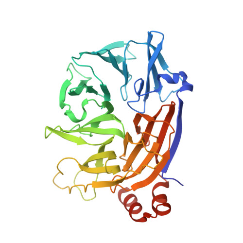
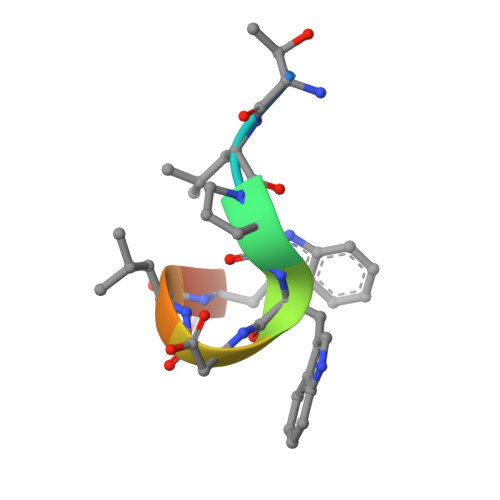
Two distinct interaction motifs in amphiphysin bind two independent sites on the clathrin terminal domain beta-propeller.

Miele, A.E.Watson, P.J.Evans, P.R.Traub, L.M.Owen, D.J.

(2004) Nat Struct Mol Biol 11: 242-248

  • DOI: https://doi.org/10.1038/nsmb736
  • Primary Citation of Related Structures:  
    1UTC

  • PubMed Abstract: 

    During the assembly of clathrin-coated vesicles, many peripheral membrane proteins, including the amphiphysins, use LLDLD-type clathrin-box motifs to interact with the N-terminal beta-propeller domain (TD) of clathrin. The 2.3 A-resolution structure of the clathrin TD in complex with a TLPWDLWTT peptide from amphiphysin 1 delineates a second clathrin-binding motif, PWXXW (the W box), that binds at a site on the TD remote from the clathrin box-binding site. The presence of both sequence motifs within the unstructured region of the amphiphysins allows them to bind more tightly to free TDs than do other endocytic proteins that contain only clathrin-box motifs. This property, along with the propensity of the N-terminal BAR domain to bind curved membranes, will preferentially localize amphiphysin and its partner, dynamin, to the periphery of invaginated clathrin lattices.


  • Organizational Affiliation

    MRC Laboratory of Molecular Biology, Hills Road, Cambridge CB2 2QH, UK.


Macromolecules
Find similar proteins by:  (by identity cutoff)  |  3D Structure
Entity ID: 1
MoleculeChains Sequence LengthOrganismDetailsImage
CLATHRIN HEAVY CHAIN
A, B
363Bos taurusMutation(s): 0 
UniProt
Find proteins for P49951 (Bos taurus)
Explore P49951 
Go to UniProtKB:  P49951
Entity Groups  
Sequence Clusters30% Identity50% Identity70% Identity90% Identity95% Identity100% Identity
UniProt GroupP49951
Sequence Annotations
Expand
  • Reference Sequence

Find similar proteins by:  Sequence   |   3D Structure  

Entity ID: 2
MoleculeChains Sequence LengthOrganismDetailsImage
AMPHIPHYSINC [auth P],
D [auth Q]
9Homo sapiensMutation(s): 0 
UniProt & NIH Common Fund Data Resources
Find proteins for P49418 (Homo sapiens)
Explore P49418 
Go to UniProtKB:  P49418
PHAROS:  P49418
GTEx:  ENSG00000078053 
Entity Groups  
UniProt GroupP49418
Sequence Annotations
Expand
  • Reference Sequence
Experimental Data & Validation

Experimental Data

  • Method: X-RAY DIFFRACTION
  • Resolution: 2.30 Å
  • R-Value Free: 0.253 
  • R-Value Work: 0.194 
  • R-Value Observed: 0.197 
  • Space Group: P 41 21 2
Unit Cell:
Length ( Å )Angle ( ˚ )
a = 61.301α = 90
b = 61.301β = 90
c = 420.52γ = 90
Software Package:
Software NamePurpose
REFMACrefinement
MOSFLMdata reduction
SCALAdata scaling
AMoREphasing

Structure Validation

View Full Validation Report


Entry History 

Deposition Data

Revision History  (Full details and data files)

  • Version 1.0: 2004-02-25
    Type: Initial release
  • Version 1.1: 2011-05-08
    Changes: Version format compliance
  • Version 1.2: 2011-07-13
    Changes: Version format compliance
  • Version 1.3: 2018-02-28
    Changes: Database references, Source and taxonomy
  • Version 1.4: 2019-10-16
    Changes: Data collection, Experimental preparation, Other
  • Version 1.5: 2023-12-13
    Changes: Data collection, Database references, Refinement description