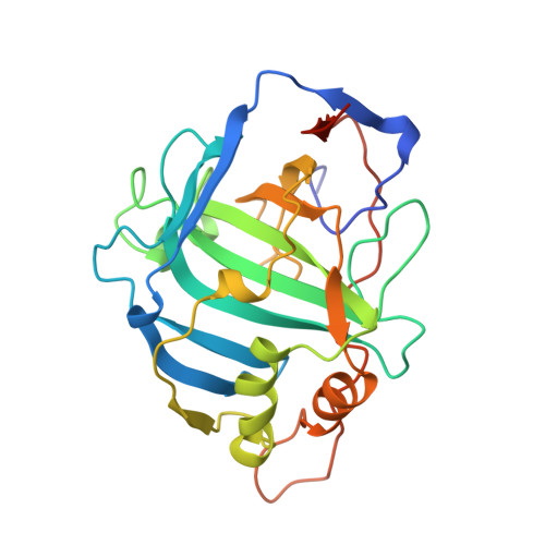
Structure-based design of an intramolecular proton transfer site in murine carbonic anhydrase V.
Heck, R.W., Boriack-Sjodin, P.A., Qian, M., Tu, C., Christianson, D.W., Laipis, P.J., Silverman, D.N.(1996) Biochemistry 35: 11605-11611
- PubMed: 8794740
- DOI: https://doi.org/10.1021/bi9608018
- Primary Citation of Related Structures:
1URT - PubMed Abstract:
Carbonic anhydrase V (CA V) is a mitochondrial enzyme that catalyzes the hydration of CO2 to produce bicarbonate and a proton. The catalytic properties of wild-type murine CA V suggest the presence of a proton shuttle residue having pKa = 9.2, the role of which is to transfer a proton from zinc-bound water to solution in the hydration direction to regenerate the zinc hydroxide form of the enzyme. Two likely candidates for shuttle residues are the tyrosines at positions 64 and 131 in the active site cavity. The crystal structure of wild-type carbonic anhydrase V [Boriack-Sjodin et al. (1995) Proc. Natl. Acad. Sci. U.S.A. 92, 10949-10953] shows that the side chain of Tyr 64 is forced into an orientation pointing away from the zinc by Phe 65, although Tyr 131 is oriented toward the zinc. We have prepared mutants of murine CA V replacing both Tyr 64 and Tyr 131 with His and Ala and investigated the proton shuttle mechanism using stopped-flow spectrophotometry and the depletion of 18O from CO2 measured by mass spectrometry. Experiments with both single and double mutations showed that neither position 64 nor position 131 was a prominent site for proton transfer. However, a double mutant of CA V containing the two replacements, Tyr 64-->His and Phe 65-->Ala, demonstrated enhanced proton transfer with an apparent pKa of 6.8 and maximal contribution to kcat of 2.2 x 10(5) s-1. In addition to the altered catalytic properties, the crystal structure of the His 64/Ala 65 double mutant strongly suggested proton transfer by His 64 after removal of the steric hindrance of Phe 65. This is the first structure-based design of an efficient proton transfer site in an enzyme.
Organizational Affiliation:
Department of Pharmacology, University of Florida, Gainesville 32610-0267, USA.