


Crystal Structure of the C47S Mutant of Human Peroxiredoxin 5
Evrard, C., Smeets, A., Knoops, B., Declercq, J.P.(2004) J Chem Crystallogr 34: 553
Experimental Data Snapshot
wwPDB Validation 3D Report Full Report

(2004) J Chem Crystallogr 34: 553
Entity ID: 1 | |||||
|---|---|---|---|---|---|
| Molecule | Chains | Sequence Length | Organism | Details | Image |
| PEROXIREDOXIN 5 | 172 | Homo sapiens | Mutation(s): 1 | 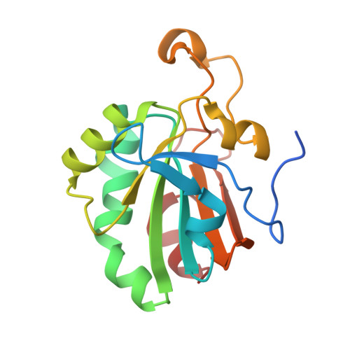 | |
UniProt & NIH Common Fund Data Resources | |||||
Find proteins for P30044 (Homo sapiens) Explore P30044 Go to UniProtKB: P30044 | |||||
PHAROS: P30044 GTEx: ENSG00000126432 | |||||
Entity Groups | |||||
| Sequence Clusters | 30% Identity50% Identity70% Identity90% Identity95% Identity100% Identity | ||||
| UniProt Group | P30044 | ||||
Sequence AnnotationsExpand | |||||
| |||||
| Ligands 2 Unique | |||||
|---|---|---|---|---|---|
| ID | Chains | Name / Formula / InChI Key | 2D Diagram | 3D Interactions | |
| BEZ Query on BEZ | B [auth A] | BENZOIC ACID C7 H6 O2 WPYMKLBDIGXBTP-UHFFFAOYSA-N |  | ||
| CL Query on CL | C [auth A], D [auth A], E [auth A] | CHLORIDE ION Cl VEXZGXHMUGYJMC-UHFFFAOYSA-M |  | ||
| Length ( Å ) | Angle ( ˚ ) |
|---|---|
| a = 65.646 | α = 90 |
| b = 65.646 | β = 90 |
| c = 122.043 | γ = 90 |
| Software Name | Purpose |
|---|---|
| REFMAC | refinement |
| DENZO | data reduction |
| SCALEPACK | data scaling |
RCSB PDB (citation) is hosted by
RCSB PDB is a member of the

