1UR5

Stabilization of a Tetrameric Malate Dehydrogenase by Introduction of a Disulfide Bridge at the Dimer/Dimer Interface


Experimental Data Snapshot

  • Method: X-RAY DIFFRACTION
  • Resolution: 1.75 Å
  • R-Value Free: 0.223 
  • R-Value Work: 0.190 
  • R-Value Observed: 0.190 

wwPDB Validation   3D Report Full Report


Ligand Structure Quality Assessment 


This is version 1.4 of the entry. See complete history


Literature

Stabilization of a Tetrameric Malate Dehydrogenase by Introduction of a Disulfide Bridge at the Dimer-Dimer Interface

Bjork, A.Dalhus, B.Mantzilas, D.Eijsink, V.G.H.Sirevag, R.

(2003) J Mol Biol 334: 811

  • DOI: https://doi.org/10.1016/j.jmb.2003.10.006
  • Primary Citation of Related Structures:  
    1UR5

  • PubMed Abstract: 

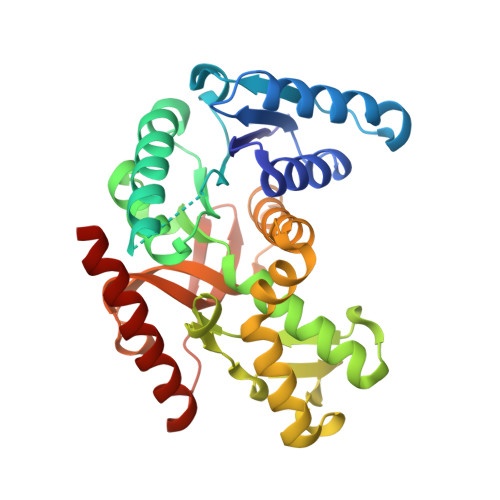
    Malate dehydrogenase (MDH) from the moderately thermophilic bacterium Chloroflexus aurantiacus (CaMDH) is a tetrameric enzyme, while MDHs from mesophilic organisms usually are dimers. To investigate the potential contribution of the extra dimer-dimer interface in CaMDH with respect to thermal stability, we have engineered an intersubunit disulfide bridge designed to strengthen dimer-dimer interactions. The resulting mutant (T187C, containing two 187-187 disulfide bridges in the tetramer) showed a 200-fold increase in half-life at 75 degrees C and an increase of 15 deg. C in apparent melting temperature compared to the wild-type. The crystal structure of the mutant (solved at 1.75 A resolution) was essentially identical with that of the wild-type, with the exception of the added inter-dimer disulfide bridge and the loss of an aromatic intra-dimer contact. Remarkably, the mutant and the wild-type had similar temperature optima and activities at their temperature optima, thus providing a clear case of uncoupling of thermal stability and thermoactivity. The results show that tetramerization may contribute to MDH stability to an extent that depends strongly on the number of stabilizing interactions in the dimer-dimer interface.


  • Organizational Affiliation

    Department of Biology, University of Oslo, PO Box 1031 Blindern, N-0316 Oslo, Norway. alexandra.bjork@bio.uio.no


Macromolecules
Find similar proteins by:  (by identity cutoff)  |  3D Structure
Entity ID: 1
MoleculeChains Sequence LengthOrganismDetailsImage
MALATE DEHYDROGENASEA,
B [auth C]
309Chloroflexus aurantiacusMutation(s): 1 
EC: 1.1.1.37
UniProt
Find proteins for P80040 (Chloroflexus aurantiacus (strain ATCC 29366 / DSM 635 / J-10-fl))
Explore P80040 
Go to UniProtKB:  P80040
Entity Groups  
Sequence Clusters30% Identity50% Identity70% Identity90% Identity95% Identity100% Identity
UniProt GroupP80040
Sequence Annotations
Expand
  • Reference Sequence
Small Molecules
Ligands 4 Unique
IDChains Name / Formula / InChI Key2D Diagram3D Interactions
NAD
Query on NAD

Download Ideal Coordinates CCD File 
C [auth A],
I [auth C]
NICOTINAMIDE-ADENINE-DINUCLEOTIDE
C21 H27 N7 O14 P2
BAWFJGJZGIEFAR-NNYOXOHSSA-N
CD
Query on CD

Download Ideal Coordinates CCD File 
D [auth A]
E [auth A]
F [auth A]
J [auth C]
K [auth C]
D [auth A],
E [auth A],
F [auth A],
J [auth C],
K [auth C],
L [auth C]
CADMIUM ION
Cd
WLZRMCYVCSSEQC-UHFFFAOYSA-N
CL
Query on CL

Download Ideal Coordinates CCD File 
G [auth A]CHLORIDE ION
Cl
VEXZGXHMUGYJMC-UHFFFAOYSA-M
NA
Query on NA

Download Ideal Coordinates CCD File 
H [auth A],
M [auth C]
SODIUM ION
Na
FKNQFGJONOIPTF-UHFFFAOYSA-N
Experimental Data & Validation

Experimental Data

  • Method: X-RAY DIFFRACTION
  • Resolution: 1.75 Å
  • R-Value Free: 0.223 
  • R-Value Work: 0.190 
  • R-Value Observed: 0.190 
  • Space Group: P 31 2 1
Unit Cell:
Length ( Å )Angle ( ˚ )
a = 105.575α = 90
b = 105.575β = 90
c = 102.531γ = 120
Software Package:
Software NamePurpose
CNSrefinement
DENZOdata reduction
SCALEPACKdata scaling
CNSphasing

Structure Validation

View Full Validation Report


Ligand Structure Quality Assessment 


Entry History 

Deposition Data

Revision History  (Full details and data files)

  • Version 1.0: 2003-11-05
    Type: Initial release
  • Version 1.1: 2011-05-08
    Changes: Version format compliance
  • Version 1.2: 2011-07-13
    Changes: Version format compliance
  • Version 1.3: 2018-02-07
    Changes: Source and taxonomy
  • Version 1.4: 2023-12-13
    Changes: Data collection, Database references, Derived calculations, Other, Refinement description