1UMU

STRUCTURE DETERMINATION OF UMUD' BY MAD PHASING OF THE SELENOMETHIONYL PROTEIN


Experimental Data Snapshot

  • Method: X-RAY DIFFRACTION
  • Resolution: 2.50 Å
  • R-Value Free: 0.303 
  • R-Value Work: 0.207 
  • R-Value Observed: 0.207 

wwPDB Validation   3D Report Full Report


This is version 1.2 of the entry. See complete history


Literature

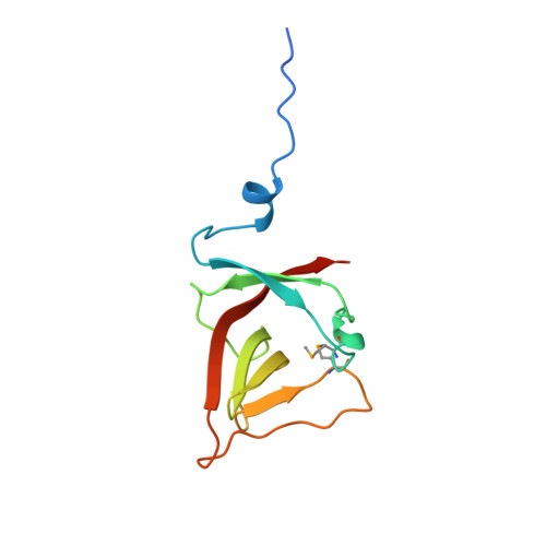
Structure of the UmuD' protein and its regulation in response to DNA damage.

Peat, T.S.Frank, E.G.McDonald, J.P.Levine, A.S.Woodgate, R.Hendrickson, W.A.

(1996) Nature 380: 727-730

  • DOI: https://doi.org/10.1038/380727a0
  • Primary Citation of Related Structures:  
    1UMU

  • PubMed Abstract: 

    For life to be sustained, mistakes in DNA repair must be tolerated when damage obscures the genetic information. In bacteria such as Escherichia coli, DNA damage elicits the well regulated 'SOS response'. For the extreme case of damage that cannot be repaired by conventional enzymes, there are proteins that allow the replication of DNA through such lesions, but with a reduction in the fidelity of replication. Essential proteins in this mutagenic process are RecA, DNA polymerase III, UmuD, UmuD' and UmuC (umu: UV mutagenesis). Regulation of this response involves a RecA-mediated self-cleavage of UmuD to produce UmuD'. To understand this system in more detail, we have determined the crystal structure of the E. coli UmuD' mutagenesis protein at 2.5 A resolution. Globular heads folded in an unusual Beta-structure associate to form molecular dimers, and extended amino-terminal tails associate to produce crystallized filaments. The structure provides insight into the mechanism of the self-cleavage reaction that UmuD-like proteins undergo as part of the global SOS response.


  • Organizational Affiliation

    Deparment of Biochemistry and Molecular Biophysics, Columbia University, NY 10032, USA.


Macromolecules
Find similar proteins by:  (by identity cutoff)  |  3D Structure
Entity ID: 1
MoleculeChains Sequence LengthOrganismDetailsImage
UMUD'
A, B
116Escherichia coliMutation(s): 0 
EC: 3.4.21
UniProt
Find proteins for P0AG11 (Escherichia coli (strain K12))
Explore P0AG11 
Go to UniProtKB:  P0AG11
Entity Groups  
Sequence Clusters30% Identity50% Identity70% Identity90% Identity95% Identity100% Identity
UniProt GroupP0AG11
Sequence Annotations
Expand
  • Reference Sequence
Small Molecules
Modified Residues  1 Unique
IDChains TypeFormula2D DiagramParent
MSE
Query on MSE
A, B
L-PEPTIDE LINKINGC5 H11 N O2 SeMET
Experimental Data & Validation

Experimental Data

  • Method: X-RAY DIFFRACTION
  • Resolution: 2.50 Å
  • R-Value Free: 0.303 
  • R-Value Work: 0.207 
  • R-Value Observed: 0.207 
  • Space Group: P 41 21 2
Unit Cell:
Length ( Å )Angle ( ˚ )
a = 52.8α = 90
b = 52.8β = 90
c = 160.1γ = 90
Software Package:
Software NamePurpose
MADSYSphasing
X-PLORmodel building
X-PLORrefinement
DENZOdata reduction
CCP4data scaling
X-PLORphasing

Structure Validation

View Full Validation Report


Entry History 

Deposition Data

Revision History  (Full details and data files)

  • Version 1.0: 1996-08-01
    Type: Initial release
  • Version 1.1: 2008-03-24
    Changes: Version format compliance
  • Version 1.2: 2011-07-13
    Changes: Version format compliance