1UK0

Crystal structure of catalytic domain of human poly(ADP-ribose) polymerase with a novel inhibitor


Experimental Data Snapshot

  • Method: X-RAY DIFFRACTION
  • Resolution: 3.00 Å
  • R-Value Free: 0.246 
  • R-Value Work: 0.219 

wwPDB Validation   3D Report Full Report


Ligand Structure Quality Assessment 


This is version 1.3 of the entry. See complete history


Literature

Inhibitor-induced structural change of the active site of human poly(ADP-ribose) polymerase.

Kinoshita, T.Nakanishi, I.Warizaya, M.Iwashita, A.Kido, Y.Hattori, K.Fujii, T.

(2004) FEBS Lett 556: 43-46

  • DOI: https://doi.org/10.1016/s0014-5793(03)01362-0
  • Primary Citation of Related Structures:  
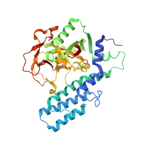
    1UK0

  • PubMed Abstract: 

    The crystal structure of human recombinant poly(ADP-ribose) polymerase (PARP) complexed with a potent inhibitor, FR257517, was solved at 3.0 A resolution. The fluorophenyl part of the inhibitor induces an amazing conformational change in the active site of PARP by motion of the side chain of the amino acid, Arg878, which forms the bottom of the active site. Consequently, a corn-shaped hydrophobic subsite, which consists of the side chains of Leu769, Ile879, Pro881, and the methylene chain of Arg878, newly emerges from the well-known active site.


  • Organizational Affiliation

    Exploratory Research Laboratories, Fujisawa Pharmaceutical Co. Ltd., 5-2-3, Tokodai, Tsukuba, 300-2698, Ibaraki, Japan. takayoshi_kinoshita@po.fujisawa.co.jp


Macromolecules
Find similar proteins by:  (by identity cutoff)  |  3D Structure
Entity ID: 1
MoleculeChains Sequence LengthOrganismDetailsImage
Poly [ADP-ribose] polymerase-1
A, B
350Homo sapiensMutation(s): 0 
EC: 2.4.2.30
UniProt & NIH Common Fund Data Resources
Find proteins for P09874 (Homo sapiens)
Explore P09874 
Go to UniProtKB:  P09874
PHAROS:  P09874
GTEx:  ENSG00000143799 
Entity Groups  
Sequence Clusters30% Identity50% Identity70% Identity90% Identity95% Identity100% Identity
UniProt GroupP09874
Sequence Annotations
Expand
  • Reference Sequence
Small Molecules
Ligands 1 Unique
IDChains Name / Formula / InChI Key2D Diagram3D Interactions
FRM
Query on FRM

Download Ideal Coordinates CCD File 
C [auth A],
D [auth B]
2-{3-[4-(4-FLUOROPHENYL)-3,6-DIHYDRO-1(2H)-PYRIDINYL]PROPYL}-8-METHYL-4(3H)-QUINAZOLINONE
C23 H24 F N3 O
LOFDUAJQRUYHBR-UHFFFAOYSA-N
Binding Affinity Annotations 
IDSourceBinding Affinity
FRM BindingDB:  1UK0 IC50: min: 8, max: 16 (nM) from 2 assay(s)
PDBBind:  1UK0 IC50: 8 (nM) from 1 assay(s)
Experimental Data & Validation

Experimental Data

  • Method: X-RAY DIFFRACTION
  • Resolution: 3.00 Å
  • R-Value Free: 0.246 
  • R-Value Work: 0.219 
  • Space Group: C 1 2 1
Unit Cell:
Length ( Å )Angle ( ˚ )
a = 179.82α = 90
b = 53.55β = 114.4
c = 92.01γ = 90
Software Package:
Software NamePurpose
DENZOdata reduction
SCALEPACKdata scaling
AMoREphasing
CNXrefinement

Structure Validation

View Full Validation Report


Ligand Structure Quality Assessment 


Entry History 

Deposition Data

Revision History  (Full details and data files)

  • Version 1.0: 2004-01-27
    Type: Initial release
  • Version 1.1: 2008-04-27
    Changes: Version format compliance
  • Version 1.2: 2011-07-13
    Changes: Version format compliance
  • Version 1.3: 2023-12-27
    Changes: Data collection, Database references, Derived calculations