1UJN

Crystal structure of dehydroquinate synthase from Thermus thermophilus HB8


Experimental Data Snapshot

  • Method: X-RAY DIFFRACTION
  • Resolution: 1.80 Å
  • R-Value Free: 0.236 
  • R-Value Work: 0.202 
  • R-Value Observed: 0.203 

wwPDB Validation   3D Report Full Report


This is version 1.3 of the entry. See complete history


Literature

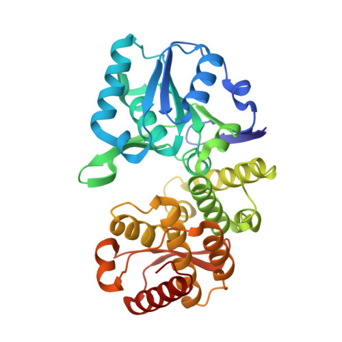
Crystal structure of dehydroquinate synthase from Thermus thermophilus HB8 showing functional importance of the dimeric state.

Sugahara, M.Nodake, Y.Sugahara, M.Kunishima, N.

(2005) Proteins 58: 249-252


Macromolecules
Find similar proteins by:  (by identity cutoff)  |  3D Structure
Entity ID: 1
MoleculeChains Sequence LengthOrganismDetailsImage
dehydroquinate synthase
A, B
348Thermus thermophilus HB8Mutation(s): 0 
EC: 4.2.3.4
UniProt
Find proteins for P83703 (Thermus thermophilus)
Explore P83703 
Go to UniProtKB:  P83703
Entity Groups  
Sequence Clusters30% Identity50% Identity70% Identity90% Identity95% Identity100% Identity
UniProt GroupP83703
Sequence Annotations
Expand
  • Reference Sequence
Experimental Data & Validation

Experimental Data

  • Method: X-RAY DIFFRACTION
  • Resolution: 1.80 Å
  • R-Value Free: 0.236 
  • R-Value Work: 0.202 
  • R-Value Observed: 0.203 
  • Space Group: P 1 21 1
Unit Cell:
Length ( Å )Angle ( ˚ )
a = 63.403α = 90
b = 71.787β = 99.44
c = 71.636γ = 90
Software Package:
Software NamePurpose
HKL-2000data collection
SCALEPACKdata scaling
AMoREphasing
CNSrefinement
HKL-2000data reduction

Structure Validation

View Full Validation Report


Entry History 

Deposition Data

Revision History  (Full details and data files)

  • Version 1.0: 2003-09-02
    Type: Initial release
  • Version 1.1: 2008-04-27
    Changes: Version format compliance
  • Version 1.2: 2011-07-13
    Changes: Source and taxonomy, Version format compliance
  • Version 1.3: 2023-10-25
    Changes: Data collection, Database references, Refinement description