1UCU

R-type straight flagellar filament made of full-length flagellin


Experimental Data Snapshot

  • Method: ELECTRON MICROSCOPY
  • Resolution: 4.00 Å
  • Aggregation State: FILAMENT 
  • Reconstruction Method: HELICAL 

wwPDB Validation   3D Report Full Report


This is version 1.3 of the entry. See complete history


Literature

Complete atomic model of the bacterial flagellar filament by electron cryomicroscopy

Yonekura, K.Maki-Yonekura, S.Namba, K.

(2003) Nature 424: 643-650

  • DOI: https://doi.org/10.1038/nature01830
  • Primary Citation of Related Structures:  
    1UCU

  • PubMed Abstract: 

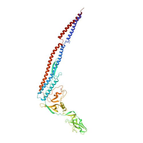
    The bacterial flagellar filament is a helical propeller for bacterial locomotion. It is a helical assembly of a single protein, flagellin, and its tubular structure is formed by 11 protofilaments in two distinct conformations, L- and R-type, for supercoiling. The X-ray crystal structure of a flagellin fragment lacking about 100 terminal residues revealed the protofilament structure, but the full filament structure is still essential for understanding the mechanism of supercoiling and polymerization. Here we report a complete atomic model of the R-type filament by electron cryomicroscopy. A density map obtained from image data up to 4 A resolution shows the feature of alpha-helical backbone and some large side chains. The atomic model built on the map reveals intricate molecular packing and an alpha-helical coiled coil formed by the terminal chains in the inner core of the filament, with its intersubunit hydrophobic interactions having an important role in stabilizing the filament.


  • Organizational Affiliation

    Protonic NanoMachine Project, ERATO, JST.


Macromolecules
Find similar proteins by:  (by identity cutoff)  |  3D Structure
Entity ID: 1
MoleculeChains Sequence LengthOrganismDetailsImage
phase 1 Flagellin494Salmonella enterica subsp. enterica serovar TyphimuriumMutation(s): 0 
UniProt
Find proteins for P06179 (Salmonella typhimurium (strain LT2 / SGSC1412 / ATCC 700720))
Explore P06179 
Go to UniProtKB:  P06179
Entity Groups  
Sequence Clusters30% Identity50% Identity70% Identity90% Identity95% Identity100% Identity
UniProt GroupP06179
Sequence Annotations
Expand
  • Reference Sequence
Experimental Data & Validation

Experimental Data

  • Method: ELECTRON MICROSCOPY
  • Resolution: 4.00 Å
  • Aggregation State: FILAMENT 
  • Reconstruction Method: HELICAL 

Structure Validation

View Full Validation Report


Entry History 

Deposition Data

Revision History  (Full details and data files)

  • Version 1.0: 2003-08-12
    Type: Initial release
  • Version 1.1: 2008-04-27
    Changes: Version format compliance
  • Version 1.2: 2011-07-13
    Changes: Version format compliance
  • Version 1.3: 2024-03-13
    Changes: Author supporting evidence, Data collection, Database references, Experimental preparation, Refinement description, Structure summary