1U8X

CRYSTAL STRUCTURE OF GLVA FROM BACILLUS SUBTILIS, A METAL-REQUIRING, NAD-DEPENDENT 6-PHOSPHO-ALPHA-GLUCOSIDASE


Experimental Data Snapshot

  • Method: X-RAY DIFFRACTION
  • Resolution: 2.05 Å
  • R-Value Free: 0.221 
  • R-Value Work: 0.195 
  • R-Value Observed: 0.197 

wwPDB Validation   3D Report Full Report


Ligand Structure Quality Assessment 


This is version 1.4 of the entry. See complete history


Literature

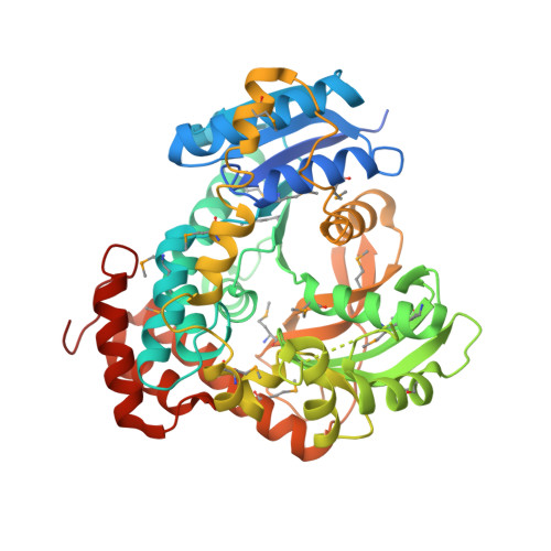
Novel Catalytic Mechanism of Glycoside Hydrolysis Based on the Structure of an NAD(+)/Mn(2+)-Dependent Phospho-alpha-Glucosidase from Bacillus subtilis.

Rajan, S.S.Yang, X.Collart, F.Yip, V.L.Withers, S.G.Varrot, A.Thompson, J.Davies, G.J.Anderson, W.F.

(2004) Structure 12: 1619-1629

  • DOI: https://doi.org/10.1016/j.str.2004.06.020
  • Primary Citation of Related Structures:  
    1U8X

  • PubMed Abstract: 

    GlvA, a 6-phospho-alpha-glucosidase from Bacillus subtilis, catalyzes the hydrolysis of maltose-6'-phosphate and belongs to glycoside hydrolase family GH4. GH4 enzymes are unique in their requirement for NAD(H) and a divalent metal for activity. We have determined the crystal structure of GlvA in complex with its ligands to 2.05 A resolution. Analyses of the active site architecture, in conjunction with mechanistic studies and precedent from the nucleotide diphosphate hexose dehydratases and other systems, suggest a novel mechanism of glycoside hydrolysis by GlvA that involves both the NAD(H) and the metal.


  • Organizational Affiliation

    Molecular Pharmacology and Biological Chemistry, Feinberg School of Medicine, Northwestern University, Chicago, IL 60611, USA.


Macromolecules
Find similar proteins by:  (by identity cutoff)  |  3D Structure
Entity ID: 1
MoleculeChains Sequence LengthOrganismDetailsImage
Maltose-6'-phosphate glucosidaseA [auth X]472Bacillus subtilisMutation(s): 17 
Gene Names: glvAglvGglv-1
EC: 3.2.1.122
UniProt
Find proteins for P54716 (Bacillus subtilis (strain 168))
Explore P54716 
Go to UniProtKB:  P54716
Entity Groups  
Sequence Clusters30% Identity50% Identity70% Identity90% Identity95% Identity100% Identity
UniProt GroupP54716
Sequence Annotations
Expand
  • Reference Sequence
Small Molecules
Ligands 3 Unique
IDChains Name / Formula / InChI Key2D Diagram3D Interactions
NAD
Query on NAD

Download Ideal Coordinates CCD File 
D [auth X]NICOTINAMIDE-ADENINE-DINUCLEOTIDE
C21 H27 N7 O14 P2
BAWFJGJZGIEFAR-NNYOXOHSSA-N
G6P
Query on G6P

Download Ideal Coordinates CCD File 
B [auth X]6-O-phosphono-alpha-D-glucopyranose
C6 H13 O9 P
NBSCHQHZLSJFNQ-DVKNGEFBSA-N
MN
Query on MN

Download Ideal Coordinates CCD File 
C [auth X]MANGANESE (II) ION
Mn
WAEMQWOKJMHJLA-UHFFFAOYSA-N
Modified Residues  1 Unique
IDChains TypeFormula2D DiagramParent
MSE
Query on MSE
A [auth X]L-PEPTIDE LINKINGC5 H11 N O2 SeMET
Experimental Data & Validation

Experimental Data

  • Method: X-RAY DIFFRACTION
  • Resolution: 2.05 Å
  • R-Value Free: 0.221 
  • R-Value Work: 0.195 
  • R-Value Observed: 0.197 
  • Space Group: I 2 2 2
Unit Cell:
Length ( Å )Angle ( ˚ )
a = 82.604α = 90
b = 102.017β = 90
c = 144.748γ = 90
Software Package:
Software NamePurpose
DENZOdata reduction
SCALEPACKdata scaling
SOLVEphasing
REFMACrefinement

Structure Validation

View Full Validation Report


Ligand Structure Quality Assessment 


Entry History 

Deposition Data

Revision History  (Full details and data files)

  • Version 1.0: 2004-08-24
    Type: Initial release
  • Version 1.1: 2008-04-30
    Changes: Version format compliance
  • Version 1.2: 2011-07-13
    Changes: Advisory, Version format compliance
  • Version 1.3: 2020-07-29
    Type: Remediation
    Reason: Carbohydrate remediation
    Changes: Advisory, Data collection, Database references, Derived calculations, Structure summary
  • Version 1.4: 2024-04-03
    Changes: Data collection, Database references, Refinement description, Structure summary