1U70

Understanding the Role of Leu22 Variants in Methotrexate Resistance: Comparison of Wild-type and Leu22Arg Variant Mouse and Human Dihydrofolate Reductase


Experimental Data Snapshot

  • Method: X-RAY DIFFRACTION
  • Resolution: 2.50 Å
  • R-Value Free: 0.225 
  • R-Value Work: 0.183 
  • R-Value Observed: 0.225 

wwPDB Validation   3D Report Full Report


Ligand Structure Quality Assessment 


This is version 1.5 of the entry. See complete history


Literature

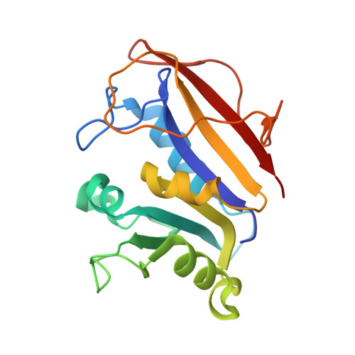
Understanding the role of Leu22 variants in methotrexate resistance: comparison of wild-type and Leu22Arg variant mouse and human dihydrofolate reductase ternary crystal complexes with methotrexate and NADPH.

Cody, V.Luft, J.R.Pangborn, W.

(2005) Acta Crystallogr D Biol Crystallogr 61: 147-155

  • DOI: https://doi.org/10.1107/S0907444904030422
  • Primary Citation of Related Structures:  
    1U70, 1U71, 1U72

  • PubMed Abstract: 

    Structural data are reported to 2.5 A resolution for the first full analysis of the methotrexate-resistant Leu22Arg (L22R) variant of mouse dihydrofolate reductase (mDHFR) crystallized as a ternary complex with methotrexate (MTX) and the cofactor NADPH. These results are compared with the MTX and NADPH ternary complexes of L22R human DHFR (hDHFR) and those of mouse and human wild-type DHFR enzymes. The conformation of mDHFR Arg22 is such that it makes hydrogen-bonding contacts with Asp21, Trp24 and a structural water molecule, observations which were not made in the L22R hDHFR ternary complex. These data show that there is little difference between the structures of the wild type and L22R variant for either mouse or human DHFR; however, there are significant differences between the species. Comparison of these structures reveals that the active site of mDHFR is larger than that in the hDHFR structure. In mDHFR, the position of MTX is shifted 0.6 A toward helix C (residues 59-65), which in turn is shifted 1.2 A away from the active site relative to that observed in the hDHFR ternary complexes. In the L22R variant mDHFR structure, MTX makes shorter contacts to the conserved residues Ile7, Val115 and Tyr121 than in the L22R variant human DHFR structure. These contacts are comparable in both wild-type enzymes. The unexpected results from this comparison of the mouse and human DHFR complexes bound with the same ligand and cofactor illustrate the importance of detailed study of several species of enzyme, even when there is a high sequence homology between them. These data suggest that the differences in binding interactions of the L22R variant are in agreement with the weaker binding affinity for MTX in the variant enzymes; the larger size of the binding site in mDHFR supports the observation that the binding affinity of MTX for L22R mDHFR is significantly weaker than that of the L22R hDHFR enzyme.


  • Organizational Affiliation

    Hauptman-Woodward Medical Research Institute, 73 High Street, Buffalo, NY 14203, USA. cody@hwi.buffalo.edu


Macromolecules
Find similar proteins by:  (by identity cutoff)  |  3D Structure
Entity ID: 1
MoleculeChains Sequence LengthOrganismDetailsImage
Dihydrofolate reductase186Mus musculusMutation(s): 1 
Gene Names: Dhfr
EC: 1.5.1.3
UniProt & NIH Common Fund Data Resources
Find proteins for P00375 (Mus musculus)
Explore P00375 
Go to UniProtKB:  P00375
IMPC:  MGI:94890
Entity Groups  
Sequence Clusters30% Identity50% Identity70% Identity90% Identity95% Identity100% Identity
UniProt GroupP00375
Sequence Annotations
Expand
  • Reference Sequence
Small Molecules
Ligands 2 Unique
IDChains Name / Formula / InChI Key2D Diagram3D Interactions
NDP
Query on NDP

Download Ideal Coordinates CCD File 
C [auth A]NADPH DIHYDRO-NICOTINAMIDE-ADENINE-DINUCLEOTIDE PHOSPHATE
C21 H30 N7 O17 P3
ACFIXJIJDZMPPO-NNYOXOHSSA-N
MTX
Query on MTX

Download Ideal Coordinates CCD File 
B [auth A]METHOTREXATE
C20 H22 N8 O5
FBOZXECLQNJBKD-ZDUSSCGKSA-N
Binding Affinity Annotations 
IDSourceBinding Affinity
MTX Binding MOAD:  1U70 Ki: 230 (nM) from 1 assay(s)
BindingDB:  1U70 Ki: min: 3.40e-4, max: 3.4 (nM) from 14 assay(s)
Kd: 0.19 (nM) from 1 assay(s)
IC50: min: 0.6, max: 1500 (nM) from 61 assay(s)
EC50: min: 12.5, max: 13.3 (nM) from 2 assay(s)
Experimental Data & Validation

Experimental Data

  • Method: X-RAY DIFFRACTION
  • Resolution: 2.50 Å
  • R-Value Free: 0.225 
  • R-Value Work: 0.183 
  • R-Value Observed: 0.225 
  • Space Group: P 1 21 1
Unit Cell:
Length ( Å )Angle ( ˚ )
a = 42.016α = 90
b = 61.536β = 116.69
c = 43.572γ = 90
Software Package:
Software NamePurpose
DENZOdata reduction
SCALEPACKdata scaling
PROTEINmodel building
PROFFTrefinement
PROTEINphasing

Structure Validation

View Full Validation Report


Ligand Structure Quality Assessment 


Entry History 

Deposition Data

  • Released Date: 2005-02-01 
  • Deposition Author(s): Cody, V.

Revision History  (Full details and data files)

  • Version 1.0: 2005-02-01
    Type: Initial release
  • Version 1.1: 2008-04-30
    Changes: Version format compliance
  • Version 1.2: 2011-07-13
    Changes: Version format compliance
  • Version 1.3: 2018-02-14
    Changes: Experimental preparation
  • Version 1.4: 2021-10-20
    Changes: Database references, Derived calculations
  • Version 1.5: 2024-02-14
    Changes: Data collection