Independent Movement, Dimerization and Stability of Tandem Repeats of Chicken Brain alpha-Spectrin
Kusunoki, H., Minasov, G., Macdonald, R.I., Mondragon, A.(2004) J Mol Biol 344: 495-511
- PubMed: 15522301
- DOI: https://doi.org/10.1016/j.jmb.2004.09.019
- Primary Citation of Related Structures:
1U4Q, 1U5P - PubMed Abstract:
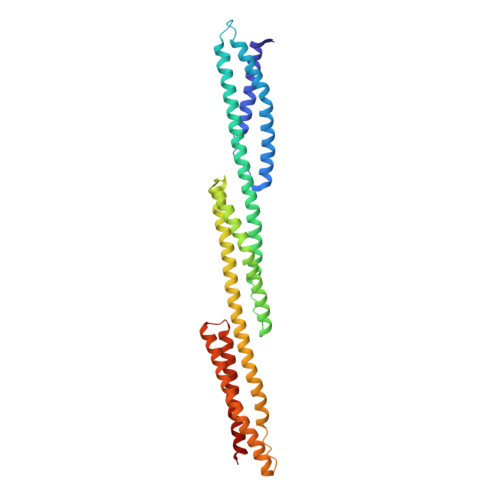
Previous X-ray crystal structures have shown that linkers of five amino acid residues connecting pairs of chicken brain alpha-spectrin and human erythroid beta-spectrin repeats can undergo bending without losing their alpha-helical structure. To test whether bending at one linker can influence bending at an adjacent linker, the structures of two and three repeat fragments of chicken brain alpha-spectrin have been determined by X-ray crystallography. The structure of the three-repeat fragment clearly shows that bending at one linker can occur independently of bending at an adjacent linker. This observation increases the possible trajectories of modeled chains of spectrin repeats. Furthermore, the three-repeat molecule crystallized as an antiparallel dimer with a significantly smaller buried interfacial area than that of alpha-actinin, a spectrin-related molecule, but large enough and of a type indicating biological specificity. Comparison of the structures of the spectrin and alpha-actinin dimers supports weak association of the former, which could not be detected by analytical ultracentrifugation, versus strong association of the latter, which has been observed by others. To correlate features of the structure with solution properties and to test a previous model of stable spectrin and dystrophin repeats, the number of inter-helical interactions in each repeat of several spectrin structures were counted and compared to their thermal stabilities. Inter-helical interactions, but not all interactions, increased in parallel with measured thermal stabilities of each repeat and in agreement with the thermal stabilities of two and three repeats and also partial repeats of spectrin.
Organizational Affiliation:
Department of Biochemistry, Molecular Biology and Cell Biology, Northwestern University, 2205 Tech Drive, Evanston, IL 60208, USA.