1U0D

Y33H Mutant of Homing endonuclease I-CreI


Experimental Data Snapshot

  • Method: X-RAY DIFFRACTION
  • Resolution: 2.90 Å
  • R-Value Free: 0.257 
  • R-Value Work: 0.217 
  • R-Value Observed: 0.242 

wwPDB Validation   3D Report Full Report


This is version 1.3 of the entry. See complete history


Literature

Isolation and characterization of new homing endonuclease specificities at individual target site positions.

Sussman, D.Chadsey, M.Fauce, S.Engel, A.Bruett, A.Monnat, R.Stoddard, B.L.Seligman, L.M.

(2004) J Mol Biol 342: 31-41

  • DOI: https://doi.org/10.1016/j.jmb.2004.07.031
  • Primary Citation of Related Structures:  
    1U0C, 1U0D

  • PubMed Abstract: 

    Homing endonucleases are highly specific DNA endonucleases, encoded within mobile introns or inteins, that induce targeted recombination, double-strand repair and gene conversion of their cognate target sites. Due to their biological function and high level of target specificity, these enzymes are under intense investigation as tools for gene targeting. These studies require that naturally occurring enzymes be redesigned to recognize novel target sites. Here, we report studies in which the homodimeric LAGLIDADG homing endonuclease I-CreI is altered at individual side-chains corresponding to contact points to distinct base-pairs in its target site. The resulting enzyme constructs drive specific elimination of selected DNA targets in vivo and display shifted specificities of DNA binding and cleavage in vitro. Crystal structures of two of these constructs demonstrate that substitution of individual side-chain/DNA contact patterns can occur with almost no structural deformation or rearrangement of the surrounding complex, facilitating an isolated, modular redesign strategy for homing endonuclease activity and specificity.


  • Organizational Affiliation

    Division of Basic Sciences, Fred Hutchinson Cancer Research Center, 1100 Fairview Ave. N. A3-025 Seattle, WA 98109, USA.


Macromolecules

Find similar proteins by:  (by identity cutoff)  |  3D Structure
Entity ID: 3
MoleculeChains Sequence LengthOrganismDetailsImage
DNA endonuclease I-CreIC [auth A],
D [auth B]
163Chlamydomonas reinhardtiiMutation(s): 5 
EC: 3.1
UniProt
Find proteins for P05725 (Chlamydomonas reinhardtii)
Explore P05725 
Go to UniProtKB:  P05725
Entity Groups  
Sequence Clusters30% Identity50% Identity70% Identity90% Identity95% Identity100% Identity
UniProt GroupP05725
Sequence Annotations
Expand
  • Reference Sequence

Find similar nucleic acids by:  Sequence   |   3D Structure  

Entity ID: 1
MoleculeChains LengthOrganismImage
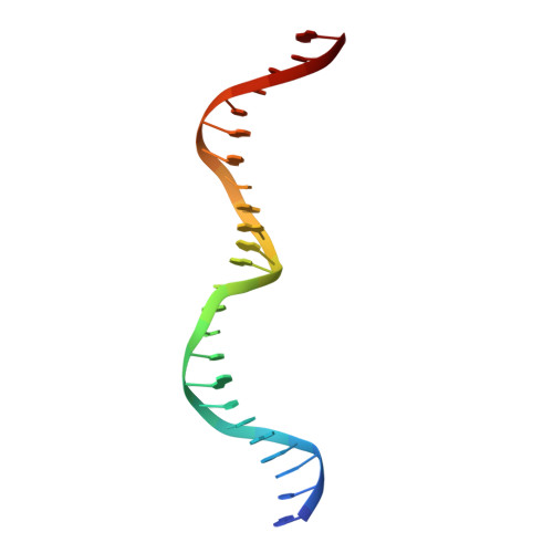
5'-D(*GP*CP*GP*AP*AP*AP*CP*GP*TP*CP*GP*TP*GP*AP*GP*AP*CP*AP*GP*TP*TP*CP*CP*G)-3'A [auth C]24N/A
Sequence Annotations
Expand
  • Reference Sequence

Find similar nucleic acids by:  Sequence   |   3D Structure  

Entity ID: 2
MoleculeChains LengthOrganismImage
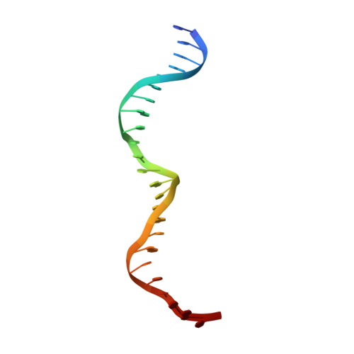
5'-D(*CP*GP*GP*AP*AP*CP*TP*GP*TP*CP*TP*CP*AP*CP*GP*AP*CP*GP*TP*TP*TP*CP*GP*C)-3'B [auth D]24N/A
Sequence Annotations
Expand
  • Reference Sequence
Small Molecules
Binding Affinity Annotations 
IDSourceBinding Affinity
DNA PDBBind:  1U0D Kd: 2 (nM) from 1 assay(s)
Experimental Data & Validation

Experimental Data

  • Method: X-RAY DIFFRACTION
  • Resolution: 2.90 Å
  • R-Value Free: 0.257 
  • R-Value Work: 0.217 
  • R-Value Observed: 0.242 
  • Space Group: P 1
Unit Cell:
Length ( Å )Angle ( ˚ )
a = 43.838α = 90.28
b = 70.331β = 89.94
c = 88.016γ = 98.78
Software Package:
Software NamePurpose
CNSrefinement

Structure Validation

View Full Validation Report


Entry History 

Deposition Data

Revision History  (Full details and data files)

  • Version 1.0: 2004-11-02
    Type: Initial release
  • Version 1.1: 2008-04-30
    Changes: Version format compliance
  • Version 1.2: 2011-07-13
    Changes: Version format compliance
  • Version 1.3: 2024-02-14
    Changes: Data collection, Database references