1TZS

Crystal Structure of an activation intermediate of Cathepsin E


Experimental Data Snapshot

  • Method: X-RAY DIFFRACTION
  • Resolution: 2.35 Å
  • R-Value Free: 0.238 
  • R-Value Work: 0.198 

wwPDB Validation   3D Report Full Report


This is version 1.3 of the entry. See complete history


Literature

Crystal structure of an activation intermediate of cathepsin e

Ostermann, N.Gerhartz, B.Worpenberg, S.Trappe, J.Eder, J.

(2004) J Mol Biol 342: 889-899

  • DOI: https://doi.org/10.1016/j.jmb.2004.07.073
  • Primary Citation of Related Structures:  
    1TZS

  • PubMed Abstract: 

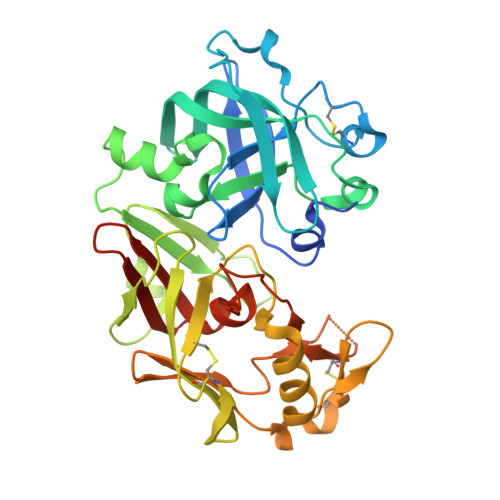
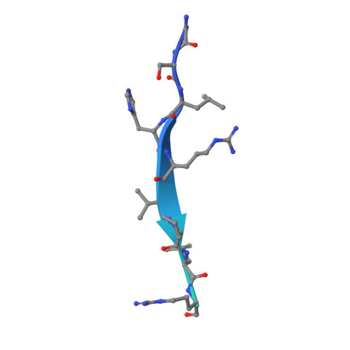
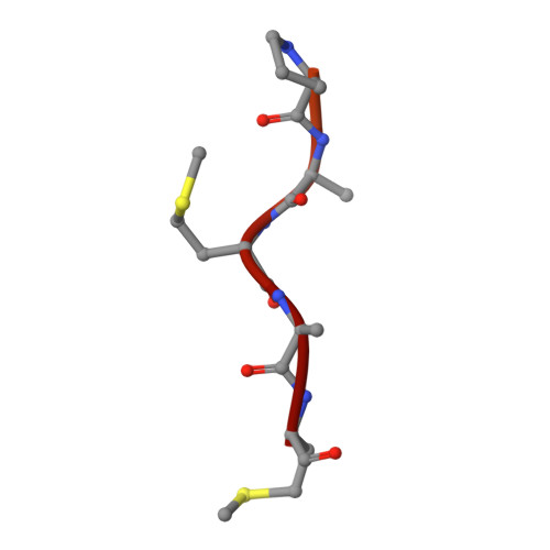
    Cathepsin E is an intracellular, non-lysosomal aspartic protease expressed in a variety of cells and tissues. The protease has proposed physiological roles in antigen presentation by the MHC class II system, in the biogenesis of the vasoconstrictor peptide endothelin, and in neurodegeneration associated with brain ischemia and aging. Cathepsin E is the only A1 aspartic protease that exists as a homodimer with a disulfide bridge linking the two monomers. Like many other aspartic proteases, it is synthesized as a zymogen which is catalytically inactive towards its natural substrates at neutral pH and which auto-activates in an acidic environment. Here we report the crystal structure of an activation intermediate of human cathepsin E at 2.35A resolution. The overall structure follows the general fold of aspartic proteases of the A1 family, and the intermediate shares many features with the intermediate 2 on the proposed activation pathway of aspartic proteases like pepsin C and cathepsin D. The pro-sequence is cleaved from the protease and remains stably associated with the mature enzyme by forming the outermost sixth strand of the interdomain beta-sheet. However, different from these other aspartic proteases the pro-sequence of cathepsin E remains intact after cleavage from the mature enzyme. In addition, the active site of cathepsin E in the crystal is occupied by N-terminal amino acid residues of the mature protease in the non-primed binding site and by an artificial N-terminal extension of the pro-sequence from a neighboring molecule in the primed site. The crystal structure of the cathepsin E/pro-sequence complex, therefore, provides further insight into the activation mechanism of aspartic proteases.


  • Organizational Affiliation

    Protease Platform, Novartis Institutes for BioMedical Research, CH-4002 Basel, Switzerland.


Macromolecules
Find similar proteins by:  (by identity cutoff)  |  3D Structure
Entity ID: 1
MoleculeChains Sequence LengthOrganismDetailsImage
Cathepsin E351Homo sapiensMutation(s): 0 
EC: 3.4.23.34
UniProt & NIH Common Fund Data Resources
Find proteins for P14091 (Homo sapiens)
Explore P14091 
Go to UniProtKB:  P14091
PHAROS:  P14091
GTEx:  ENSG00000196188 
Entity Groups  
Sequence Clusters30% Identity50% Identity70% Identity90% Identity95% Identity100% Identity
UniProt GroupP14091
Sequence Annotations
Expand
  • Reference Sequence
Find similar proteins by:  (by identity cutoff)  |  3D Structure
Entity ID: 2
MoleculeChains Sequence LengthOrganismDetailsImage
activation peptide from Cathepsin EB [auth P]35Homo sapiensMutation(s): 0 
EC: 3.4.23.34
UniProt & NIH Common Fund Data Resources
Find proteins for P14091 (Homo sapiens)
Explore P14091 
Go to UniProtKB:  P14091
PHAROS:  P14091
GTEx:  ENSG00000196188 
Entity Groups  
Sequence Clusters30% Identity50% Identity70% Identity90% Identity95% Identity100% Identity
UniProt GroupP14091
Sequence Annotations
Expand
  • Reference Sequence

Find similar proteins by:  Sequence   |   3D Structure  

Entity ID: 3
MoleculeChains Sequence LengthOrganismDetailsImage
23-mer peptide from PelB-IgG kappa light chain fusion proteinC [auth X]23N/AMutation(s): 0 
Entity Groups  
Sequence Clusters30% Identity50% Identity70% Identity90% Identity95% Identity100% Identity
Sequence Annotations
Expand
  • Reference Sequence
Experimental Data & Validation

Experimental Data

  • Method: X-RAY DIFFRACTION
  • Resolution: 2.35 Å
  • R-Value Free: 0.238 
  • R-Value Work: 0.198 
  • Space Group: P 41 21 2
Unit Cell:
Length ( Å )Angle ( ˚ )
a = 61.321α = 90
b = 61.321β = 90
c = 207.8γ = 90
Software Package:
Software NamePurpose
CNXrefinement
MOSFLMdata reduction
CCP4data scaling
MOLREPphasing

Structure Validation

View Full Validation Report


Entry History 

Deposition Data

Revision History  (Full details and data files)

  • Version 1.0: 2005-07-12
    Type: Initial release
  • Version 1.1: 2008-04-30
    Changes: Version format compliance
  • Version 1.2: 2011-07-13
    Changes: Version format compliance
  • Version 1.3: 2023-10-25
    Changes: Data collection, Database references, Refinement description