1TW8

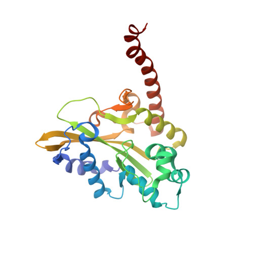
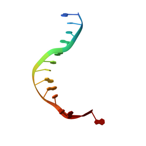
HincII bound to Ca2+ and cognate DNA GTCGAC


Experimental Data Snapshot

  • Method: X-RAY DIFFRACTION
  • Resolution: 2.80 Å
  • R-Value Free: 0.286 
  • R-Value Work: 0.180 

wwPDB Validation   3D Report Full Report


This is version 1.5 of the entry. See complete history


Literature

Ca2+ binding in the active site of HincII: implications for the catalytic mechanism

Etzkorn, C.Horton, N.C.

(2004) Biochemistry 43: 13256-13270

  • DOI: https://doi.org/10.1021/bi0490082
  • Primary Citation of Related Structures:  
    1TW8, 1TX3

  • PubMed Abstract: 

    The 2.8 A crystal structure of the type II restriction endonuclease HincII bound to Ca(2+) and cognate DNA containing GTCGAC is presented. The DNA is uncleaved, and one calcium ion is bound per active site, in a position previously described as site I in the related blunt cutting type II restriction endonuclease EcoRV [Horton, N. C., Newberry, K. J., and Perona, J. J. (1998) Proc. Natl. Acad. Sci. U.S.A. 95 (23), 13489-13494], as well as that found in other related enzymes. Unlike the site I metal in EcoRV, but similar to that of PvuII, NgoMIV, BamHI, BglII, and BglI, the observed calcium cation is directly ligated to the pro-S(p) oxygen of the scissile phosphate. A calcium ion-ligated water molecule is well positioned to act as the nucleophile in the phosphodiester bond cleavage reaction, and is within hydrogen bonding distance of the conserved active site lysine (Lys 129), as well as the pro-R(p) oxygen of the phosphate group 3' of the scissile phosphate, suggesting possible roles for these groups in the catalytic mechanism. Kinetic data consistent with an important role for the 3'-phosphate group in DNA cleavage by HincII are presented. The previously observed sodium ion [Horton, N. C., Dorner, L. F., and Perona, J. J. (2002) Nat. Struct. Biol. 9, 42-47] persists in the active sites of the Ca(2+)-bound structure; however, kinetic data show little effect on the single-turnover rate of DNA cleavage in the absence of Na(+) ions.


  • Organizational Affiliation

    Department of Biochemistry and Molecular Biophysics, University of Arizona, Tucson, Arizona 85721, USA.


Macromolecules

Find similar proteins by:  (by identity cutoff)  |  3D Structure
Entity ID: 2
MoleculeChains Sequence LengthOrganismDetailsImage
Hinc II endonucleaseE [auth A],
F [auth B],
G [auth C],
H [auth D]
257Haemophilus influenzaeMutation(s): 0 
Gene Names: Hinc II
EC: 3.1.21.4
UniProt
Find proteins for P17743 (Haemophilus influenzae)
Explore P17743 
Go to UniProtKB:  P17743
Entity Groups  
Sequence Clusters30% Identity50% Identity70% Identity90% Identity95% Identity100% Identity
UniProt GroupP17743
Sequence Annotations
Expand
  • Reference Sequence

Find similar nucleic acids by:  Sequence   |   3D Structure  

Entity ID: 1
MoleculeChains LengthOrganismImage
5'-D(*GP*CP*CP*GP*GP*TP*CP*GP*AP*CP*CP*GP*G)-3'A [auth E],
B [auth F],
C [auth G],
D [auth H]
13N/A
Sequence Annotations
Expand
  • Reference Sequence
Experimental Data & Validation

Experimental Data

  • Method: X-RAY DIFFRACTION
  • Resolution: 2.80 Å
  • R-Value Free: 0.286 
  • R-Value Work: 0.180 
  • Space Group: I 21 21 21
Unit Cell:
Length ( Å )Angle ( ˚ )
a = 66.945α = 90
b = 177.18β = 90
c = 254.89γ = 90
Software Package:
Software NamePurpose
MOSFLMdata reduction
SCALAdata scaling
TRUNCATEdata reduction
CNSrefinement
CCP4data scaling
TRUNCATEdata scaling
CNSphasing

Structure Validation

View Full Validation Report


Entry History 

Deposition Data

Revision History  (Full details and data files)

  • Version 1.0: 2004-08-10
    Type: Initial release
  • Version 1.1: 2008-04-30
    Changes: Version format compliance
  • Version 1.2: 2011-07-13
    Changes: Version format compliance
  • Version 1.3: 2012-06-06
    Changes: Database references, Structure summary
  • Version 1.4: 2017-10-11
    Changes: Refinement description
  • Version 1.5: 2023-08-23
    Changes: Data collection, Database references, Derived calculations, Refinement description