Structure of the complex between trypanosomal triosephosphate isomerase and N-hydroxy-4-phosphono-butanamide: binding at the active site despite an "open" flexible loop conformation.
Verlinde, C.L., Witmans, C.J., Pijning, T., Kalk, K.H., Hol, W.G., Callens, M., Opperdoes, F.R.(1992) Protein Sci 1: 1578-1584
- PubMed: 1304889
- DOI: https://doi.org/10.1002/pro.5560011205
- Primary Citation of Related Structures:
1TSI - PubMed Abstract:
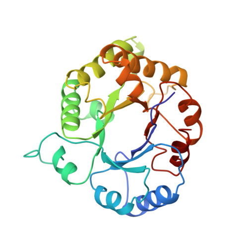
The structure of triosephosphate isomerase from Trypanosoma brucei complexed with the competitive inhibitor N-hydroxy-4-phosphono-butanamide was determined by X-ray crystallography to a resolution of 2.84 A. Full occupancy binding of the inhibitor is observed only at one of the active sites of the homodimeric enzyme where the flexible loop is locked in a completely open conformation by crystal contacts. There is evidence that the inhibitor also binds to the second active site of the enzyme, but with low occupancy. The hydroxamyl group of the inhibitor forms hydrogen bonds to the side chains of Asn 11, Lys 13, and His 95, whereas each of its three methylene units is involved in nonpolar interactions with the side chain of the flexible loop residue Ile 172. Interactions between the hydroxamyl and the catalytic base Glu 167 are absent. The binding of this phosphonate inhibitor exhibits three unusual features: (1) the flexible loop is open, in contrast with the binding mode observed in eight other complexes between triosephosphate isomerase and various phosphate and phosphonate compounds; (2) compared with these complexes the present structure reveals a 1.5-A shift of the anion-binding site; (3) this is the first phosphonate inhibitor that is not forced by the enzyme into an eclipsed conformation about the P-CH2 bond. The results are discussed with respect to an ongoing drug design project aimed at the selective inhibition of glycolytic enzymes of T. brucei.
Organizational Affiliation:
BIOSON Research Institute, University of Groningen, The Netherlands.