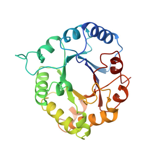
The structural basis for pseudoreversion of the H95N lesion by the secondary S96P mutation in triosephosphate isomerase.
Komives, E.A., Lougheed, J.C., Zhang, Z., Sugio, S., Narayana, N., Xuong, N.H., Petsko, G.A., Ringe, D.(1996) Biochemistry 35: 15474-15484
- PubMed: 8952501
- DOI: https://doi.org/10.1021/bi961556v
- Primary Citation of Related Structures:
1TPU, 1TPV - PubMed Abstract:
The structural basis for the 3000-fold decrease in catalytic efficiency of the H95N mutant chicken triosephosphate isomerase and the 60-fold regain of catalytic efficiency in the double mutant, H95N.S96P, have been analyzed. The results from a combination of X-ray crystallography and Fourier transform infrared spectroscopy experiments indicate that the predominant defect in the H95N mutant isomerase appears to be its inability to bind the substrate in a coplanar, cis conformation. The structures of each mutant isomerase were determined from X-ray crystallography of the complex of phosphoglycolohydroxamate (PGH), an intermediate analog with the isomerase, and each was solved to a resolution of 1.9 A. The PGH appeared to be in two different conformations in which the enediol-mimicking atoms, O2-N2-C1-O1, of the PGH were not coplanar. No density was observed that would correspond to the coplanar conformation. Two bands are observed for the dihydroxyacetone phosphate carbonyl in the H95N mutant FTIR spectrum, and these can be explained if the O1 of DHAP, like the O1 of PGH in the crystal structure, is in two different positions. Two ordered water molecules are located between O1 of PGH and N delta of N95. Comparison of the structure of the pseudorevertant, H95N.S96P with that for the H95N single mutant, shows that S96P mutation causes the double mutant to regain the ability to bind PGH predominantly in the coplanar, cis conformation. Electron density for a single ordered water molecule bridging the N95 amide side chain and the O2 of PGH is observed, but the density was weak, perhaps indicating that the water molecule is somewhat disordered. Whether or not a water molecule is hydrogen bonded to O2 of PGH may explain the two carbonyl stretching frequencies observed for the GAP carbonyl. Together, the crystal structures and the FTIR data allow a complete explanation of the catalytic properties of these two mutant isomerases.
Organizational Affiliation:
Department of Chemistry and Biochemistry, University of California, San Diego, La Jolla 92093-0601, USA.