1TOH

TYROSINE HYDROXYLASE CATALYTIC AND TETRAMERIZATION DOMAINS FROM RAT


Experimental Data Snapshot

  • Method: X-RAY DIFFRACTION
  • Resolution: 2.30 Å
  • R-Value Free: 0.268 
  • R-Value Work: 0.211 
  • R-Value Observed: 0.211 

wwPDB Validation   3D Report Full Report


This is version 1.3 of the entry. See complete history


Literature

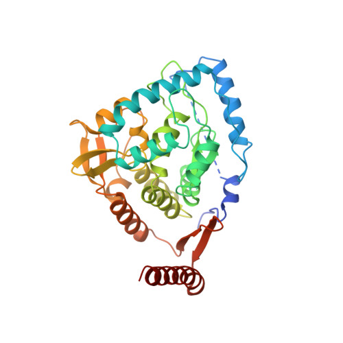
Crystal structure of tyrosine hydroxylase at 2.3 A and its implications for inherited neurodegenerative diseases.

Goodwill, K.E.Sabatier, C.Marks, C.Raag, R.Fitzpatrick, P.F.Stevens, R.C.

(1997) Nat Struct Biol 4: 578-585

  • DOI: https://doi.org/10.1038/nsb0797-578
  • Primary Citation of Related Structures:  
    1TOH

  • PubMed Abstract: 

    Tyrosine hydroxylase (TyrOH) catalyzes the conversion of tyrosine to L-DOPA, the rate-limiting step in the biosynthesis of the catecholamines dopamine, adrenaline, and noradrenaline. TyrOH is highly homologous in terms of both protein sequence and catalytic mechanism to phenylalanine hydroxylase (PheOH) and tryptophan hydroxylase (TrpOH). The crystal structure of the catalytic and tetramerization domains of TyrOH reveals a novel alpha-helical basket holding the catalytic iron and a 40 A long anti-parallel coiled coil which forms the core of the tetramer. The catalytic iron is located 10 A below the enzyme surface in a 17 A deep active site pocket and is coordinated by the conserved residues His 331, His 336 and Glu 376. The structure provides a rationale for the effect of point mutations in TyrOH that cause L-DOPA responsive parkinsonism and Segawa's syndrome. The location of 112 different point mutations in PheOH that lead to phenylketonuria (PKU) are predicted based on the TyrOH structure.


  • Organizational Affiliation

    Department of Chemistry, University of California, Berkeley 94720, USA.


Macromolecules
Find similar proteins by:  (by identity cutoff)  |  3D Structure
Entity ID: 1
MoleculeChains Sequence LengthOrganismDetailsImage
TYROSINE HYDROXYLASE343Rattus norvegicusMutation(s): 0 
EC: 1.14.16.2
UniProt
Find proteins for P04177 (Rattus norvegicus)
Explore P04177 
Go to UniProtKB:  P04177
Entity Groups  
Sequence Clusters30% Identity50% Identity70% Identity90% Identity95% Identity100% Identity
UniProt GroupP04177
Sequence Annotations
Expand
  • Reference Sequence
Small Molecules
Ligands 1 Unique
IDChains Name / Formula / InChI Key2D Diagram3D Interactions
FE
Query on FE

Download Ideal Coordinates CCD File 
B [auth A]FE (III) ION
Fe
VTLYFUHAOXGGBS-UHFFFAOYSA-N
Experimental Data & Validation

Experimental Data

  • Method: X-RAY DIFFRACTION
  • Resolution: 2.30 Å
  • R-Value Free: 0.268 
  • R-Value Work: 0.211 
  • R-Value Observed: 0.211 
  • Space Group: F 2 2 2
Unit Cell:
Length ( Å )Angle ( ˚ )
a = 189.323α = 90
b = 148.335β = 90
c = 57.975γ = 90
Software Package:
Software NamePurpose
CNSrefinement
DENZOdata reduction
SCALEPACKdata scaling
CNSphasing

Structure Validation

View Full Validation Report


Entry History 

Deposition Data

Revision History  (Full details and data files)

  • Version 1.0: 1998-06-03
    Type: Initial release
  • Version 1.1: 2008-03-24
    Changes: Version format compliance
  • Version 1.2: 2011-07-13
    Changes: Derived calculations, Version format compliance
  • Version 1.3: 2024-02-14
    Changes: Data collection, Database references, Derived calculations