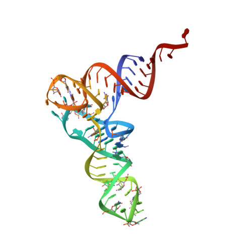
Crystallographic and biochemical investigation of the lead(II)-catalyzed hydrolysis of yeast phenylalanine tRNA.
Brown, R.S., Dewan, J.C., Klug, A.(1985) Biochemistry 24: 4785-4801
- PubMed: 3907691
- DOI: https://doi.org/10.1021/bi00339a012
- Primary Citation of Related Structures:
1TN1, 1TN2 - PubMed Abstract:
X-ray diffraction data from monoclinic crystals of yeast tRNAPhe soaked in dilute lead(II) acetate solutions at pH 5.0 and at pH 7.4 have been collected to a resolution of 3 A, and the Pb(II) binding sites have been obtained by difference Fourier analyses. The same three Pb(II) binding sites are observed at both of these pH values. At pH 7.4 an extra peak of negative electron density appears on the difference map close to one of the Pb(II) binding sites and at the position of phosphate-18, indicating cleavage of the sugar-phosphate-chain between residues D-17 and G-18 of the tRNAPhe molecule in this derivative. Chain scission does not occur to any observable extent in the structure at pH 5.0, and we have, therefore, a picture of the reactants (at pH 5.0) and products (at pH 7.4) of this cleavage reaction. Polyacrylamide gel electrophoresis as well as sequencing experiments confirms the cleavage of the tRNAPhe molecule into one-fourth and three-fourth fragments, with the shorter fragment consisting essentially of residues G-1 through D-17 while the larger fragment contains residues G-18 through A-76. End-group analyses suggest a ribose cyclic 2',3'-phosphate at D-17 of the one-fourth fragment with a 5'-OH at G-18 of the three-fourth fragment. Cleavage of the tRNAPhe molecule does not occur in the absence of Pb(II), and the proximity of one of these metal ions to the cleavage site strongly implicates this metal ion in the cleavage reaction. Consideration of several possible mechanisms for the reaction, taking into account the biochemical and crystallographic facts presented above, suggests that the cleavage involves removal of the proton from the 2'-OH of ribose-17 by a Pb(II)-bound hydroxyl group. Subsequent nucleophilic attack of the resulting 2'-O- on the phosphorus atom of phosphate-18, presumably through a pentacoordinate phosphorus cyclic intermediate (as in the action of pancreatic ribonuclease A), results in chain scission. It cannot be decided whether the displacement, within the pentacoordinate intermediate, proceeds via an in-line or adjacent pathway, but an exploration of the likelihood of either pathway is presented. Strand cleavage at the particular site occurs fortuitously because the aquo Pb(II) ion binds at the correct distance and presumably in such a manner as to present a hydroxyl group in the correct orientation to effect the proton abstraction.(ABSTRACT TRUNCATED AT 400 WORDS)