Crystal structure of the catalytic domain of a thermophilic endocellulase.
Spezio, M., Wilson, D.B., Karplus, P.A.(1993) Biochemistry 32: 9906-9916
- PubMed: 8399160
- DOI: https://doi.org/10.1021/bi00089a006
- Primary Citation of Related Structures:
1TML - PubMed Abstract:
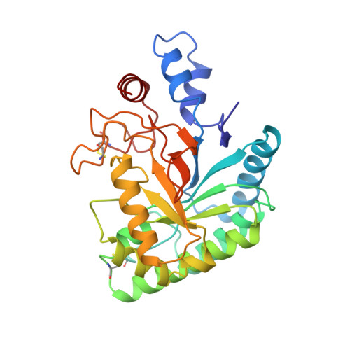
One way to improve the economic feasibility of biomass conversion is to enhance the catalytic efficiency of cellulases through protein engineering. This requires that high-resolution structures of cellulases be available. Here we present the structure of E2cd, the catalytic domain of the thermophilic endocellulase E2 from Thermomonospora fusca, as determined by X-ray crystallography. The structure was solved by multiple isomorphous replacement at 2.6-A resolution and has been refined at 1.8-A resolution to an R-value of 18.4% for all reflections between 10- and 1.8-A resolution. The fold of E2cd is based on an unusual parallel beta-barrel and is equivalent to the fold determined for the catalytic domain of cellobiohydrolase II, an exocellulase from Trichoderma reesei [Rouvinen et al. (1990) Science 249, 380-385]. The active site cleft of the enzyme, approximately 11 A deep and running the entire length of the molecule, is seen to be completely free for ligand binding in the crystal. A 2.2-A resolution analysis of crystals of E2cd complexed with cellobiose, an inhibitor, shows how cellobiose binds in the active site and interacts with several residues which line the cleft. Catalytic roles are suggested for three aspartic acid residues at the active site. A comparison of the E2cd and CBHIIcd structures reveals a large difference in their active site accessibilities and supports the hypothesis that the main difference between endo- and exocellulases is the degree to which their active sites are accessible to substrate.
Organizational Affiliation:
Section of Biochemistry, Molecular and Cell Biology, Cornell University, Ithaca, New York 14853.