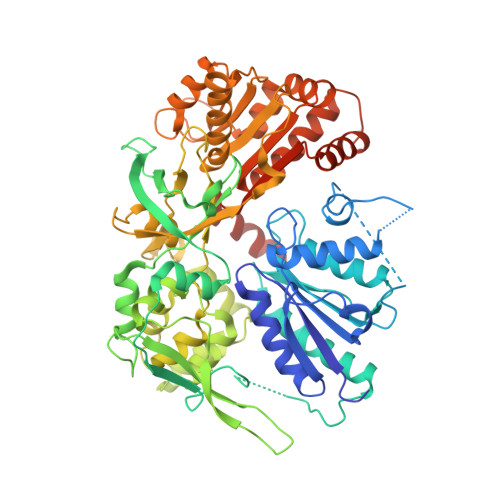
1TLL

CRYSTAL STRUCTURE OF RAT NEURONAL NITRIC-OXIDE SYNTHASE REDUCTASE MODULE AT 2.3 A RESOLUTION.


Experimental Data Snapshot

  • Method: X-RAY DIFFRACTION
  • Resolution: 2.30 Å
  • R-Value Free: 0.272 
  • R-Value Work: 0.244 
  • R-Value Observed: 0.244 

wwPDB Validation   3D Report Full Report


Ligand Structure Quality Assessment 


This is version 1.3 of the entry. See complete history


Literature

Structural basis for isozyme-specific regulation of electron transfer in nitric-oxide synthase

Garcin, E.D.Bruns, C.M.Lloyd, S.J.Hosfield, D.J.Tiso, M.Gachhui, R.Stuehr, D.J.Tainer, J.A.Getzoff, E.D.

(2004) J Biol Chem 279: 37918-37927

  • DOI: https://doi.org/10.1074/jbc.M406204200
  • Primary Citation of Related Structures:  
    1TLL

  • PubMed Abstract: 

    Three nitric-oxide synthase (NOS) isozymes play crucial, but distinct, roles in neurotransmission, vascular homeostasis, and host defense, by catalyzing Ca(2+)/calmodulin-triggered NO synthesis. Here, we address current questions regarding NOS activity and regulation by combining mutagenesis and biochemistry with crystal structure determination of a fully assembled, electron-supplying, neuronal NOS reductase dimer. By integrating these results, we structurally elucidate the unique mechanisms for isozyme-specific regulation of electron transfer in NOS. Our discovery of the autoinhibitory helix, its placement between domains, and striking similarities with canonical calmodulin-binding motifs, support new mechanisms for NOS inhibition. NADPH, isozyme-specific residue Arg(1400), and the C-terminal tail synergistically repress NOS activity by locking the FMN binding domain in an electron-accepting position. Our analyses suggest that calmodulin binding or C-terminal tail phosphorylation frees a large scale swinging motion of the entire FMN domain to deliver electrons to the catalytic module in the holoenzyme.


  • Organizational Affiliation

    Department of Molecular Biology and the Skaggs Institute for Chemical Biology, The Scripps Research Institute, La Jolla, California 92037, USA. edg@scripps.edu


Macromolecules
Find similar proteins by:  (by identity cutoff)  |  3D Structure
Entity ID: 1
MoleculeChains Sequence LengthOrganismDetailsImage
Nitric-oxide synthase, brain
A, B
688Rattus norvegicusMutation(s): 0 
Gene Names: NOS1BNOS
EC: 1.14.13.39
UniProt
Find proteins for P29476 (Rattus norvegicus)
Explore P29476 
Go to UniProtKB:  P29476
Entity Groups  
Sequence Clusters30% Identity50% Identity70% Identity90% Identity95% Identity100% Identity
UniProt GroupP29476
Sequence Annotations
Expand
  • Reference Sequence
Small Molecules
Ligands 4 Unique
IDChains Name / Formula / InChI Key2D Diagram3D Interactions
FAD
Query on FAD

Download Ideal Coordinates CCD File 
E [auth A],
I [auth B]
FLAVIN-ADENINE DINUCLEOTIDE
C27 H33 N9 O15 P2
VWWQXMAJTJZDQX-UYBVJOGSSA-N
NAP
Query on NAP

Download Ideal Coordinates CCD File 
F [auth A],
J [auth B]
NADP NICOTINAMIDE-ADENINE-DINUCLEOTIDE PHOSPHATE
C21 H28 N7 O17 P3
XJLXINKUBYWONI-NNYOXOHSSA-N
FMN
Query on FMN

Download Ideal Coordinates CCD File 
D [auth A],
H [auth B]
FLAVIN MONONUCLEOTIDE
C17 H21 N4 O9 P
FVTCRASFADXXNN-SCRDCRAPSA-N
SO3
Query on SO3

Download Ideal Coordinates CCD File 
C [auth A],
G [auth B]
SULFITE ION
O3 S
LSNNMFCWUKXFEE-UHFFFAOYSA-L
Experimental Data & Validation

Experimental Data

  • Method: X-RAY DIFFRACTION
  • Resolution: 2.30 Å
  • R-Value Free: 0.272 
  • R-Value Work: 0.244 
  • R-Value Observed: 0.244 
  • Space Group: P 1
Unit Cell:
Length ( Å )Angle ( ˚ )
a = 65.756α = 76.8
b = 69.174β = 72.07
c = 82.628γ = 67.14
Software Package:
Software NamePurpose
CNSrefinement
DENZOdata reduction
SCALEPACKdata scaling
AMoREphasing

Structure Validation

View Full Validation Report


Ligand Structure Quality Assessment 


Entry History 

Deposition Data

Revision History  (Full details and data files)

  • Version 1.0: 2004-08-31
    Type: Initial release
  • Version 1.1: 2008-04-30
    Changes: Version format compliance
  • Version 1.2: 2011-07-13
    Changes: Version format compliance
  • Version 1.3: 2023-08-23
    Changes: Data collection, Database references, Derived calculations, Refinement description