1TKY

Crystal structure of the editing domain of threonyl-tRNA synthetase complexed with seryl-3'-aminoadenosine


Experimental Data Snapshot

  • Method: X-RAY DIFFRACTION
  • Resolution: 1.48 Å
  • R-Value Free: 0.222 
  • R-Value Work: 0.201 
  • R-Value Observed: 0.203 

wwPDB Validation   3D Report Full Report


Ligand Structure Quality Assessment 


This is version 1.3 of the entry. See complete history


Literature

Achieving Error-Free Translation; The Mechanism of Proofreading of Threonyl-tRNA Synthetase at Atomic Resolution.

Dock-Bregeon, A.C.Rees, B.Torres-Larios, A.Bey, G.Caillet, J.Moras, D.

(2004) Mol Cell 16: 375-386

  • DOI: https://doi.org/10.1016/j.molcel.2004.10.002
  • Primary Citation of Related Structures:  
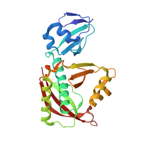
    1TJE, 1TKE, 1TKG, 1TKY

  • PubMed Abstract: 

    The fidelity of aminoacylation of tRNA(Thr) by the threonyl-tRNA synthetase (ThrRS) requires the discrimination of the cognate substrate threonine from the noncognate serine. Misacylation by serine is corrected in a proofreading or editing step. An editing site has been located 39 A away from the aminoacylation site. We report the crystal structures of this editing domain in its apo form and in complex with the serine product, and with two nonhydrolyzable analogs of potential substrates: the terminal tRNA adenosine charged with serine, and seryl adenylate. The structures show how serine is recognized, and threonine rejected, and provide the structural basis for the editing mechanism, a water-mediated hydrolysis of the mischarged tRNA. When the adenylate analog binds in the editing site, a phosphate oxygen takes the place of one of the catalytic water molecules, thereby blocking the reaction. This rules out a correction mechanism that would occur before the binding of the amino acid on the tRNA.


  • Organizational Affiliation

    IGBMC (CNRS/INSERM/Université Louis Pasteur), Laboratoire de Biologie et Génomique Structurales, 1, rue Laurent Fries, BP 10142, 67400 Illkirch, France.


Macromolecules
Find similar proteins by:  (by identity cutoff)  |  3D Structure
Entity ID: 1
MoleculeChains Sequence LengthOrganismDetailsImage
Threonyl-tRNA synthetase224Escherichia coliMutation(s): 0 
Gene Names: THRSB1719C2116SF1512S1630
EC: 6.1.1.3
UniProt
Find proteins for P0A8M3 (Escherichia coli (strain K12))
Explore P0A8M3 
Go to UniProtKB:  P0A8M3
Entity Groups  
Sequence Clusters30% Identity50% Identity70% Identity90% Identity95% Identity100% Identity
UniProt GroupP0A8M3
Sequence Annotations
Expand
  • Reference Sequence
Small Molecules
Ligands 1 Unique
IDChains Name / Formula / InChI Key2D Diagram3D Interactions
A3S
Query on A3S

Download Ideal Coordinates CCD File 
B [auth A]SERINE-3'-AMINOADENOSINE
C13 H19 N7 O5
ITDKSTILAWHDJI-AYEBZEFBSA-N
Experimental Data & Validation

Experimental Data

  • Method: X-RAY DIFFRACTION
  • Resolution: 1.48 Å
  • R-Value Free: 0.222 
  • R-Value Work: 0.201 
  • R-Value Observed: 0.203 
  • Space Group: P 21 21 21
Unit Cell:
Length ( Å )Angle ( ˚ )
a = 43.563α = 90
b = 45.429β = 90
c = 116.532γ = 90
Software Package:
Software NamePurpose
CNSrefinement
HKL-2000data reduction
SCALEPACKdata scaling
AMoREphasing

Structure Validation

View Full Validation Report


Ligand Structure Quality Assessment 


Entry History 

Deposition Data

Revision History  (Full details and data files)

  • Version 1.0: 2004-11-30
    Type: Initial release
  • Version 1.1: 2008-04-30
    Changes: Version format compliance
  • Version 1.2: 2011-07-13
    Changes: Version format compliance
  • Version 1.3: 2023-08-23
    Changes: Data collection, Database references, Derived calculations, Refinement description