Evolution of Enzymatic Activities in the Enolase Superfamily: Structure of a Substrate-Liganded Complex of the l-Ala-d/l-Glu Epimerase from Bacillus subtilis(,).
Klenchin, V.A., Schmidt, D.M., Gerlt, J.A., Rayment, I.(2004) Biochemistry 43: 10370-10378
- PubMed: 15301535
- DOI: https://doi.org/10.1021/bi049197o
- Primary Citation of Related Structures:
1TKK - PubMed Abstract:
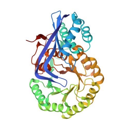
The members of the mechanistically diverse enolase superfamily share a bidomain structure formed from a (beta/alpha)7beta-barrel domain [a modified (beta/alpha)8- or TIM-barrel] and a capping domain formed from N- and C-terminal segments of the polypeptide. The active sites are located at the interface between the C-terminal ends of the beta-strands in the barrel domain and two flexible loops in the capping domain. Within this structure, the acid/base chemistry responsible for formation and stabilization of an enediolate intermediate derived from a carboxylate anion substrate and the processing of it to product is "hard-wired" by functional groups at the C-terminal ends of the beta-strands in the barrel domain; the identity of the substrate is determined in part by the identities of residues located at the end of the eighth beta-strand in the barrel domain and two mobile loops in the capping domain. On the basis of the identities of the acid/base functional groups at the ends of the beta-strands, the currently available structure-function relationships derived from functionally characterized members are often sufficient for "deciphering" the identity of the chemical reaction catalyzed by sequence-divergent members discovered in genome projects. However, insufficient structural information for liganded complexes for specifying the identity of the substrate is available. In this paper, the structure of the complex of L-Ala-L-Glu with the L-Ala-D/L-Glu epimerase from Bacillus subtilis is reported. As expected for the 1,1-proton transfer reaction catalyzed by this enzyme, the alpha-carbon of the substrate is located between Lys 162 and Lys 268 at the ends of the second and sixth beta-strands in the barrel domain. The alpha-ammonium group of the l-Ala moiety is hydrogen bonded to both Asp 321 and Asp 323 at the end of the eighth beta-strand, revealing a novel strategy for substrate recognition in the superfamily. The delta-carboxylate group of the Glu moiety is hydrogen bonded to Arg 24 in one of the flexible loops in the capping domain, thereby providing a structural explanation for the restricted substrate specificity of this epimerase [Schmidt, D. M., Hubbard, B. K., and Gerlt, J. A. (2001) Biochemistry 40, 15707-15715]. These studies provide important new information about the structural bases for substrate specificity in the enolase superfamily.
Organizational Affiliation:
Department of Biochemistry, University of Wisconsin, Madison, Wisconsin 53706, USA.