1TFF

Structure of Otubain-2


Experimental Data Snapshot

  • Method: X-RAY DIFFRACTION
  • Resolution: 2.10 Å
  • R-Value Free: 0.257 
  • R-Value Work: 0.188 
  • R-Value Observed: 0.191 

wwPDB Validation   3D Report Full Report


This is version 1.3 of the entry. See complete history


Literature

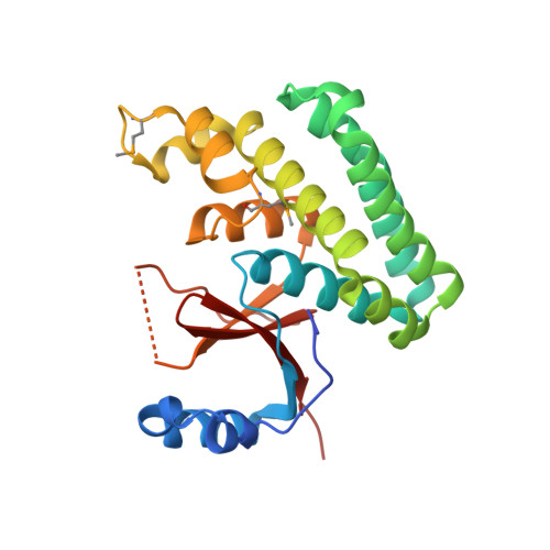
Crystal structure of human otubain 2.

Nanao, M.H.Tcherniuk, S.O.Chroboczek, J.Dideberg, O.Dessen, A.Balakirev, M.Y.

(2004) EMBO Rep 5: 783-788

  • DOI: https://doi.org/10.1038/sj.embor.7400201
  • Primary Citation of Related Structures:  
    1TFF

  • PubMed Abstract: 

    Ubiquitylation, the modification of cellular proteins by the covalent attachment of ubiquitin, is critical for diverse biological processes including cell cycle progression, signal transduction and stress response. This process can be reversed and regulated by a group of proteases called deubiquitylating enzymes (DUBs). Otubains are a recently identified family of DUBs that belong to the ovarian tumour (OTU) superfamily of proteins. Here, we report the first crystal structure of an OTU superfamily protein, otubain 2, at 2.1 A resolution and propose a model for otubain-ubiquitin binding on the basis of other DUB structures. Although otubain 2 is a member of the cysteine protease superfamily of folds, its crystal structure shows a novel fold for DUBs. Moreover, the active-site cleft is sterically occluded by a novel loop conformation resulting in an oxyanion hole, which consists uniquely of backbone amides, rather than the composite backbone/side-chain substructures seen in other DUBs and cysteine proteases. Furthermore, the residues that orient and stabilize the active-site histidine of otubain 2 are different from other cysteine proteases. This reorganization of the active-site topology provides a possible explanation for the low turnover and substrate specificity of the otubains.


  • Organizational Affiliation

    Laboratoire de Cristallographie Macromoléculaire, Institut de Biologie Structurale JP Ebel (CEA/CNRS/UJF), 41 rue Jules Horowitz, 38027 Grenoble, France.


Macromolecules
Find similar proteins by:  (by identity cutoff)  |  3D Structure
Entity ID: 1
MoleculeChains Sequence LengthOrganismDetailsImage
Ubiquitin thiolesterase protein OTUB2234Homo sapiensMutation(s): 3 
Gene Names: OTUB2OTB2OTU2C14ORF137
EC: 3.4
UniProt & NIH Common Fund Data Resources
Find proteins for Q96DC9 (Homo sapiens)
Explore Q96DC9 
Go to UniProtKB:  Q96DC9
PHAROS:  Q96DC9
GTEx:  ENSG00000089723 
Entity Groups  
Sequence Clusters30% Identity50% Identity70% Identity90% Identity95% Identity100% Identity
UniProt GroupQ96DC9
Sequence Annotations
Expand
  • Reference Sequence
Small Molecules
Modified Residues  1 Unique
IDChains TypeFormula2D DiagramParent
MSE
Query on MSE
A
L-PEPTIDE LINKINGC5 H11 N O2 SeMET
Experimental Data & Validation

Experimental Data

  • Method: X-RAY DIFFRACTION
  • Resolution: 2.10 Å
  • R-Value Free: 0.257 
  • R-Value Work: 0.188 
  • R-Value Observed: 0.191 
  • Space Group: P 21 21 21
Unit Cell:
Length ( Å )Angle ( ˚ )
a = 46.433α = 90
b = 65.401β = 90
c = 76.259γ = 90
Software Package:
Software NamePurpose
REFMACrefinement
DENZOdata reduction
SCALEPACKdata scaling
SHARPphasing

Structure Validation

View Full Validation Report


Entry History 

Deposition Data

Revision History  (Full details and data files)

  • Version 1.0: 2004-08-03
    Type: Initial release
  • Version 1.1: 2008-04-30
    Changes: Version format compliance
  • Version 1.2: 2011-07-13
    Changes: Version format compliance
  • Version 1.3: 2021-10-27
    Changes: Database references, Derived calculations