1TEZ

COMPLEX BETWEEN DNA AND THE DNA PHOTOLYASE FROM ANACYSTIS NIDULANS


Experimental Data Snapshot

  • Method: X-RAY DIFFRACTION
  • Resolution: 1.80 Å
  • R-Value Free: 0.226 
  • R-Value Work: 0.205 
  • R-Value Observed: 0.205 

wwPDB Validation   3D Report Full Report


Ligand Structure Quality Assessment 


This is version 1.4 of the entry. See complete history


Literature

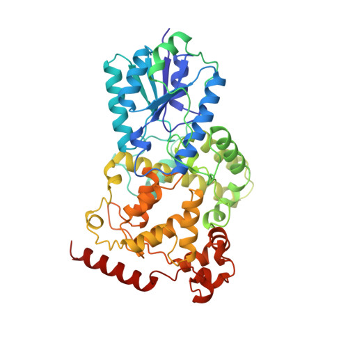
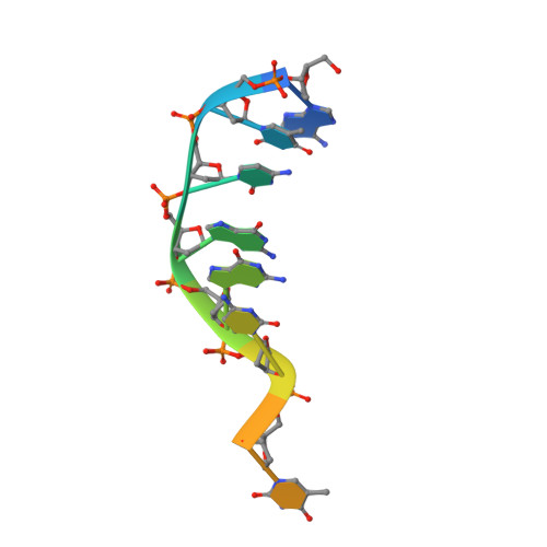
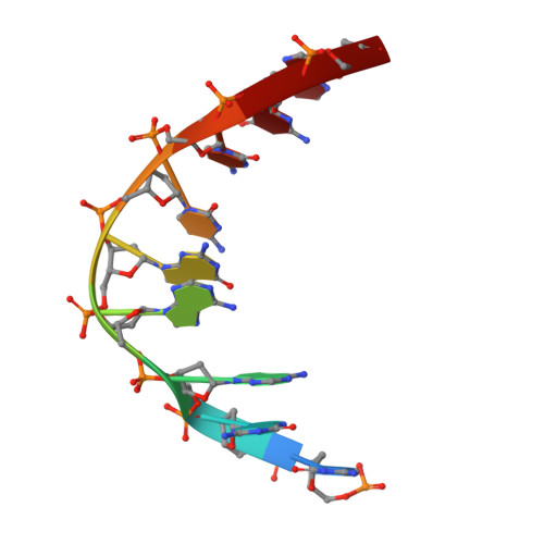
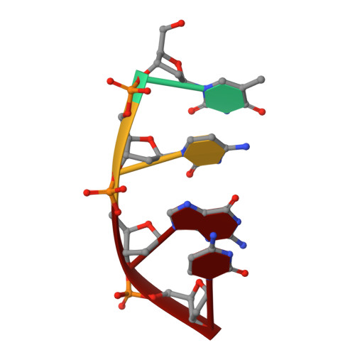
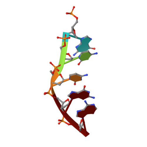
Crystal structure of a photolyase bound to a CPD-like DNA lesion after in situ repair

Mees, A.Klar, T.Gnau, P.Hennecke, U.Eker, A.P.M.Carell, T.Essen, L.-O.

(2004) Science 306: 1789-1793

  • DOI: https://doi.org/10.1126/science.1101598
  • Primary Citation of Related Structures:  
    1TEZ

  • PubMed Abstract: 

    DNA photolyases use light energy to repair DNA that comprises ultraviolet-induced lesions such as the cis-syn cyclobutane pyrimidine dimers (CPDs). Here we report the crystal structure of a DNA photolyase bound to duplex DNA that is bent by 50 degrees and comprises a synthetic CPD lesion. This CPD lesion is flipped into the active site and split there into two thymines by synchrotron radiation at 100 K. Although photolyases catalyze blue light-driven CPD cleavage only above 200 K, this structure apparently mimics a structural substate during light-driven DNA repair in which back-flipping of the thymines into duplex DNA has not yet taken place.


  • Organizational Affiliation

    Department of Chemistry and Biochemistry, Butenandt-Strasse 5-13, Ludwig Maximilians University, D-81377 Munich, Germany.


Macromolecules

Find similar proteins by:  (by identity cutoff)  |  3D Structure
Entity ID: 5
MoleculeChains Sequence LengthOrganismDetailsImage
Deoxyribodipyrimidine photolyaseI [auth A],
J [auth B],
K [auth C],
L [auth D]
474Synechococcus elongatus PCC 6301Mutation(s): 0 
Gene Names: PHR
EC: 4.1.99.3
UniProt
Find proteins for P05327 (Synechococcus sp. (strain ATCC 27144 / PCC 6301 / SAUG 1402/1))
Explore P05327 
Go to UniProtKB:  P05327
Entity Groups  
Sequence Clusters30% Identity50% Identity70% Identity90% Identity95% Identity100% Identity
UniProt GroupP05327
Sequence Annotations
Expand
  • Reference Sequence

Find similar nucleic acids by:  Sequence   |   3D Structure  

Entity ID: 1
MoleculeChains LengthOrganismImage
5'-D(*AP*TP*CP*GP*GP*CP*T*(TCP)P*CP*GP*C)-3'A [auth I],
C [auth K]
11N/A
Sequence Annotations
Expand
  • Reference Sequence

Find similar nucleic acids by:  Sequence   |   3D Structure  

Entity ID: 2
MoleculeChains LengthOrganismImage
5'-D(P*CP*GP*AP*AP*GP*CP*CP*GP*A)-3'B [auth J],
D [auth L]
9N/A
Sequence Annotations
Expand
  • Reference Sequence

Find similar nucleic acids by:  Sequence   |   3D Structure  

Entity ID: 3
MoleculeChains LengthOrganismImage
5'-D(*TP*CP*GP*C)-3'E [auth M],
G [auth O]
4N/A
Sequence Annotations
Expand
  • Reference Sequence

Find similar nucleic acids by:  Sequence   |   3D Structure  

Entity ID: 4
MoleculeChains LengthOrganismImage
5'-D(P*GP*CP*CP*GP*A)-3'F [auth N],
H [auth P]
5N/A
Sequence Annotations
Expand
  • Reference Sequence
Small Molecules
Ligands 6 Unique
IDChains Name / Formula / InChI Key2D Diagram3D Interactions
FAD
Query on FAD

Download Ideal Coordinates CCD File 
AA [auth C],
CA [auth D],
V [auth A],
Y [auth B]
FLAVIN-ADENINE DINUCLEOTIDE
C27 H33 N9 O15 P2
VWWQXMAJTJZDQX-UYBVJOGSSA-N
HDF
Query on HDF

Download Ideal Coordinates CCD File 
BA [auth C],
DA [auth D],
W [auth A],
Z [auth B]
8-HYDROXY-10-(D-RIBO-2,3,4,5-TETRAHYDROXYPENTYL)-5-DEAZAISOALLOXAZINE
C16 H17 N3 O7
AUEILLWDYUBWCM-AGIUHOORSA-N
G
Query on G

Download Ideal Coordinates CCD File 
O [auth I],
S [auth K]
GUANOSINE-5'-MONOPHOSPHATE
C10 H14 N5 O8 P
RQFCJASXJCIDSX-UUOKFMHZSA-N
C
Query on C

Download Ideal Coordinates CCD File 
N [auth I],
P [auth I],
R [auth K],
T [auth K]
CYTIDINE-5'-MONOPHOSPHATE
C9 H14 N3 O8 P
IERHLVCPSMICTF-XVFCMESISA-N
TCP
Query on TCP

Download Ideal Coordinates CCD File 
M [auth I],
Q [auth K]
5'-METHYLTHYMIDINE
C11 H16 N2 O5
KTCKNHCDUKONFQ-DJLDLDEBSA-N
MG
Query on MG

Download Ideal Coordinates CCD File 
U [auth A],
X [auth B]
MAGNESIUM ION
Mg
JLVVSXFLKOJNIY-UHFFFAOYSA-N
Experimental Data & Validation

Experimental Data

  • Method: X-RAY DIFFRACTION
  • Resolution: 1.80 Å
  • R-Value Free: 0.226 
  • R-Value Work: 0.205 
  • R-Value Observed: 0.205 
  • Space Group: C 1 2 1
Unit Cell:
Length ( Å )Angle ( ˚ )
a = 173.815α = 90
b = 88.505β = 90.11
c = 161.622γ = 90
Software Package:
Software NamePurpose
MOSFLMdata reduction
SCALAdata scaling
MOLREPphasing
CNSrefinement
CCP4data scaling

Structure Validation

View Full Validation Report


Ligand Structure Quality Assessment 


Entry History 

Deposition Data

Revision History  (Full details and data files)

  • Version 1.0: 2004-12-14
    Type: Initial release
  • Version 1.1: 2007-10-16
    Changes: Version format compliance
  • Version 1.2: 2011-07-13
    Changes: Source and taxonomy, Version format compliance
  • Version 1.3: 2018-05-30
    Changes: Advisory, Data collection, Derived calculations
  • Version 1.4: 2023-08-23
    Changes: Advisory, Data collection, Database references, Derived calculations, Refinement description