1TDG

Complex of S130G SHV-1 beta-lactamase with tazobactam


Experimental Data Snapshot

  • Method: X-RAY DIFFRACTION
  • Resolution: 1.80 Å
  • R-Value Free: 0.176 
  • R-Value Work: 0.140 
  • R-Value Observed: 0.141 

wwPDB Validation   3D Report Full Report


Ligand Structure Quality Assessment 


This is version 1.5 of the entry. See complete history


Literature

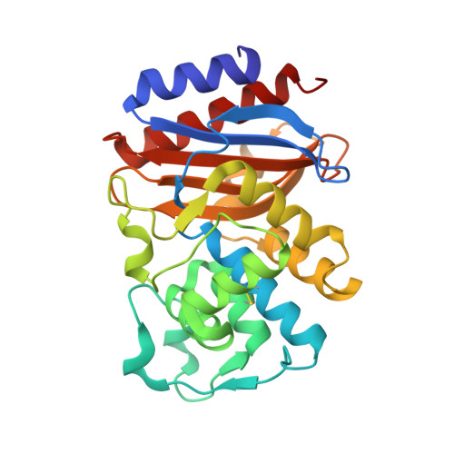
Inhibitor-resistant class A beta-lactamases: consequences of the Ser130-to-Gly mutation seen in Apo and tazobactam structures of the SHV-1 variant

Sun, T.Bethel, C.R.Bonomo, R.A.Knox, J.R.

(2004) Biochemistry 43: 14111-14117

  • DOI: https://doi.org/10.1021/bi0487903
  • Primary Citation of Related Structures:  
    1TDG, 1TDL

  • PubMed Abstract: 

    A bacterial response to the clinical use of class A beta-lactamase inhibitors such as tazobactam and clavulanic acid is the expression of variant beta-lactamases with weaker binding affinities for these mechanism-based inhibitors. Some of these inhibitor-resistant variants contain a glycine mutation at Ser130, a conserved active site residue known to be adventitiously involved in the inhibition mechanism. The crystallographic structure of a complex of tazobactam with the Ser130Gly variant of the class A SHV-1 beta-lactamase has been determined to 1.8 A resolution. Two reaction intermediates are observed. The primary intermediate is an acyclic species bound to the reactive Ser70. It is poorly primed for catalytic hydrolysis because its ester carbonyl group is completely displaced from the enzyme's oxyanion hole. A smaller fraction of the enzyme contains a Ser70-bound aldehyde resulting from hydrolytic loss of the triazoyl-sulfinyl amino acid moiety from the primary species. This first structure of a class A beta-lactamase lacking Ser130, the side chain of which functions in beta-lactam binding and possibly in catalysis, gives crystallographic evidence that the acylation step of beta-lactam turnover can occur without Ser130. Unexpectedly, the crystal structure of the uncomplexed Ser130Gly enzyme, also determined to 1.8 A resolution, shows that a critical Glu166-activated water molecule is missing from the catalytic site. Comparison of this uncomplexed variant with the wild-type structure reveals that Ser130 is required for orienting the side chain of Ser70 and ensuring the hydrogen bonding of Ser70 to both Lys73 and the catalytic water molecule.


  • Organizational Affiliation

    Department of Molecular and Cell Biology, The University of Connecticut, Storrs, Connecticut 06269-3125, USA.


Macromolecules
Find similar proteins by:  (by identity cutoff)  |  3D Structure
Entity ID: 1
MoleculeChains Sequence LengthOrganismDetailsImage
Beta-lactamase SHV-1265Klebsiella pneumoniaeMutation(s): 1 
Gene Names: bla
EC: 3.5.2.6
UniProt
Find proteins for P0AD64 (Klebsiella pneumoniae)
Explore P0AD64 
Go to UniProtKB:  P0AD64
Entity Groups  
Sequence Clusters30% Identity50% Identity70% Identity90% Identity95% Identity100% Identity
UniProt GroupP0AD64
Sequence Annotations
Expand
  • Reference Sequence
Small Molecules
Ligands 5 Unique
IDChains Name / Formula / InChI Key2D Diagram3D Interactions
MA4
Query on MA4

Download Ideal Coordinates CCD File 
B [auth A],
C [auth A]
CYCLOHEXYL-HEXYL-BETA-D-MALTOSIDE
C24 H44 O11
WUCWJXGMSXTDAV-QKMCSOCLSA-N
TBE
Query on TBE

Download Ideal Coordinates CCD File 
D [auth A]TAZOBACTAM INTERMEDIATE
C10 H14 N4 O5 S
ANZZKUOZZHRUQC-AARLMMRRSA-N
MPD
Query on MPD

Download Ideal Coordinates CCD File 
G [auth A](4S)-2-METHYL-2,4-PENTANEDIOL
C6 H14 O2
SVTBMSDMJJWYQN-YFKPBYRVSA-N
MRD
Query on MRD

Download Ideal Coordinates CCD File 
F [auth A](4R)-2-METHYLPENTANE-2,4-DIOL
C6 H14 O2
SVTBMSDMJJWYQN-RXMQYKEDSA-N
MDD
Query on MDD

Download Ideal Coordinates CCD File 
E [auth A]MALONALDEHYDE
C3 H4 O2
WSMYVTOQOOLQHP-UHFFFAOYSA-N
Experimental Data & Validation

Experimental Data

  • Method: X-RAY DIFFRACTION
  • Resolution: 1.80 Å
  • R-Value Free: 0.176 
  • R-Value Work: 0.140 
  • R-Value Observed: 0.141 
  • Space Group: P 21 21 21
Unit Cell:
Length ( Å )Angle ( ˚ )
a = 49.834α = 90
b = 55.255β = 90
c = 83.571γ = 90
Software Package:
Software NamePurpose
FRAMBOdata collection
X-GENdata reduction
EPMRphasing
CNSrefinement
X-GENdata scaling

Structure Validation

View Full Validation Report


Ligand Structure Quality Assessment 


Entry History 

Deposition Data

Revision History  (Full details and data files)

  • Version 1.0: 2004-11-23
    Type: Initial release
  • Version 1.1: 2008-04-30
    Changes: Version format compliance
  • Version 1.2: 2011-07-13
    Changes: Version format compliance
  • Version 1.3: 2017-10-11
    Changes: Refinement description
  • Version 1.4: 2021-10-27
    Changes: Database references, Derived calculations
  • Version 1.5: 2023-08-23
    Changes: Data collection, Refinement description