1TD7

Interactions of a specific non-steroidal anti-inflammatory drug (NSAID) with group I phospholipase A2 (PLA2): Crystal structure of the complex formed between PLA2 and niflumic acid at 2.5 A resolution


Experimental Data Snapshot

  • Method: X-RAY DIFFRACTION
  • Resolution: 2.50 Å
  • R-Value Free: 0.242 
  • R-Value Work: 0.193 
  • R-Value Observed: 0.195 

wwPDB Validation   3D Report Full Report


This is version 1.3 of the entry. See complete history


Literature

Non-steroidal anti-inflammatory drugs as potent inhibitors of phospholipase A2: structure of the complex of phospholipase A2 with niflumic acid at 2.5 Angstroms resolution.

Jabeen, T.Singh, N.Singh, R.K.Sharma, S.Somvanshi, R.K.Dey, S.Singh, T.P.

(2005) Acta Crystallogr D Biol Crystallogr 61: 1579-1586

  • DOI: https://doi.org/10.1107/S0907444905029604
  • Primary Citation of Related Structures:  
    1TD7

  • PubMed Abstract: 

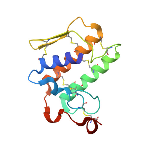
    Phospholipase A(2) (PLA(2); EC 3.1.3.4) catalyzes the first step of the production of proinflammatory compounds collectively known as eicosanoids. The binding of phospholipid substrates to PLA(2) occurs through a well formed hydrophobic channel. Surface plasmon resonance studies have shown that niflumic acid binds to Naja naja sagittifera PLA(2) with an affinity that corresponds to a dissociation constant (K(d)) of 4.3 x 10(-5) M. Binding studies of PLA(2) with niflumic acid were also carried out using a standard PLA(2) kit that gave an approximate binding constant, K(i), of 1.26 +/- 0.05 x 10(-6) M. Therefore, in order to establish the viability of PLA(2) as a potential target molecule for drug design against inflammation, arthritis and rheumatism, the three-dimensional structure of the complex of PLA(2) with the known anti-inflammatory agent niflumic acid [2-[3-(trifluoromethyl)anilino]nicotinic acid] has been determined at 2.5 Angstroms resolution. The structure of the complex has been refined to an R factor of 0.187. The structure determination reveals the presence of one niflumic acid molecule at the substrate-binding site of PLA(2). It shows that niflumic acid interacts with the important active-site residues His48 and Asp49 through two water molecules. It is observed that the niflumic acid molecule is completely buried in the substrate-binding hydrophobic channel. The conformations of the binding site in PLA(2) as well as that of niflumic acid are not altered upon binding. However, the orientation of the side chain of Trp19, which is located at the entry of the substrate-binding site, has changed from that found in the native PLA(2), indicating its familiar role.


  • Organizational Affiliation

    Department of Biophysics, All India Institute of Medical Sciences, New Delhi.


Macromolecules
Find similar proteins by:  (by identity cutoff)  |  3D Structure
Entity ID: 1
MoleculeChains Sequence LengthOrganismDetailsImage
Phospholipase A2 isoform 3119Naja sagittiferaMutation(s): 0 
EC: 3.1.1.4
UniProt
Find proteins for P60045 (Naja sagittifera)
Explore P60045 
Go to UniProtKB:  P60045
Entity Groups  
Sequence Clusters30% Identity50% Identity70% Identity90% Identity95% Identity100% Identity
UniProt GroupP60045
Sequence Annotations
Expand
  • Reference Sequence
Small Molecules
Ligands 2 Unique
IDChains Name / Formula / InChI Key2D Diagram3D Interactions
NFL
Query on NFL

Download Ideal Coordinates CCD File 
C [auth A]2-{[3-(TRIFLUOROMETHYL)PHENYL]AMINO}NICOTINIC ACID
C13 H9 F3 N2 O2
JZFPYUNJRRFVQU-UHFFFAOYSA-N
CA
Query on CA

Download Ideal Coordinates CCD File 
B [auth A]CALCIUM ION
Ca
BHPQYMZQTOCNFJ-UHFFFAOYSA-N
Binding Affinity Annotations 
IDSourceBinding Affinity
NFL PDBBind:  1TD7 Kd: 4.30e+4 (nM) from 1 assay(s)
Binding MOAD:  1TD7 Kd: 4.30e+4 (nM) from 1 assay(s)
Experimental Data & Validation

Experimental Data

  • Method: X-RAY DIFFRACTION
  • Resolution: 2.50 Å
  • R-Value Free: 0.242 
  • R-Value Work: 0.193 
  • R-Value Observed: 0.195 
  • Space Group: P 41
Unit Cell:
Length ( Å )Angle ( ˚ )
a = 42.356α = 90
b = 42.356β = 90
c = 64.958γ = 90
Software Package:
Software NamePurpose
REFMACrefinement
DENZOdata reduction
SCALEPACKdata scaling
AMoREphasing

Structure Validation

View Full Validation Report


Entry History 

Deposition Data

Revision History  (Full details and data files)

  • Version 1.0: 2004-06-08
    Type: Initial release
  • Version 1.1: 2008-04-30
    Changes: Version format compliance
  • Version 1.2: 2011-07-13
    Changes: Version format compliance
  • Version 1.3: 2023-10-25
    Changes: Data collection, Database references, Derived calculations, Refinement description