Structures for the Potential Drug Target Purine Nucleoside Phosphorylase from Schistosoma mansoni Causal Agent of Schistosomiasis.
Pereira, H.D., Franco, G.R., Cleasby, A., Garratt, R.C.(2005) J Mol Biol 353: 584-599
- PubMed: 16182308
- DOI: https://doi.org/10.1016/j.jmb.2005.08.045
- Primary Citation of Related Structures:
1TCU, 1TCV, 1TD1 - PubMed Abstract:
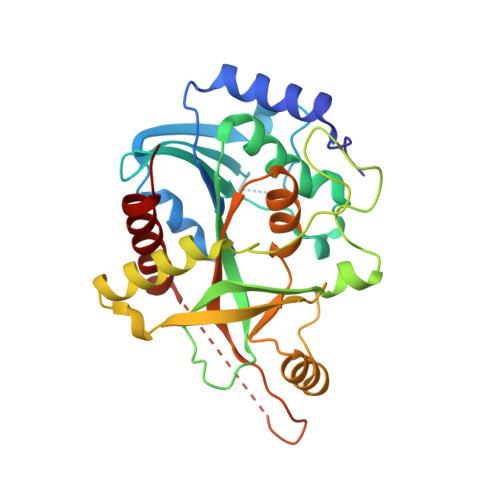
Despite the availability of effective chemotherapy, schistosomiasis continues to be one of the major parasitic infections to affect the human population worldwide. Currently, little is known of the structural biology of the parasites that are responsible for the disease and few attempts have been made to develop second generation drugs, which may become essential if resistance to those currently available becomes an issue. Here, we describe three crystal structures for the enzyme purine nucleoside phosphorylase (PNP) from Schistosoma mansoni, a component of the purine salvage pathway. PNP is known to be essential for the recovery of purine bases and nucleosides in schistosomes, due to an absence of the enzymes for de novo synthesis, making it a sensitive point in the parasite's metabolism. In all three structures reported here, acetate occupies part of the base-binding site and is directly bound to the conserved glutamic acid at position 203. One of the structures presents the crystallization additive sulfobetaine 195 (NDSB195) occupying simultaneously the ribose and phosphate binding sites, whilst a second presents only phosphate in the latter. The observation of sulfobetaine specifically bound to the protein active site was unexpected and is unique to this structure as far as we are aware. Considerable flexibility is observed in the active site, principally due to variable structural disorder in the regions centered on residues 64 and 260. This conformational plasticity extends to the way in which both NDSB195 and phosphate bind to the individual monomers of the trimeric structure reported here. Differences between the parasite and human enzymes are limited principally to the base-binding site, where the substitution of V245 in the mammalian enzymes by S247 introduces additional hydrogen bonding potential to the site. This is satisfied in the structures described here by a water molecule whose presence is normally observed only in complexes with 6-oxopurines. Residue Y202, which replaces F200 in human PNP, is able to reach over the ribose-binding site to interact with H259 and is predicted to form an additional hydrogen bond with the 5' hydroxyl of nucleoside substrates.
Organizational Affiliation:
Centro de Biotecnologia Molecular Estrutural, Instituto de Física de São Carlos, Universidade de São Paulo, CP 369, CEP 13560-970, São Carlos-SP, Brazil.