1TBQ

CRYSTAL STRUCTURE OF INSECT DERIVED DOUBLE DOMAIN KAZAL INHIBITOR RHODNIIN IN COMPLEX WITH THROMBIN


Experimental Data Snapshot

  • Method: X-RAY DIFFRACTION
  • Resolution: 3.10 Å
  • R-Value Work: 0.175 
  • R-Value Observed: 0.175 

wwPDB Validation   3D Report Full Report


This is version 1.2 of the entry. See complete history


Literature

Two heads are better than one: crystal structure of the insect derived double domain Kazal inhibitor rhodniin in complex with thrombin.

van de Locht, A.Lamba, D.Bauer, M.Huber, R.Friedrich, T.Kroger, B.Hoffken, W.Bode, W.

(1995) EMBO J 14: 5149-5157

  • DOI: https://doi.org/10.1002/j.1460-2075.1995.tb00199.x
  • Primary Citation of Related Structures:  
    1TBQ, 1TBR

  • PubMed Abstract: 

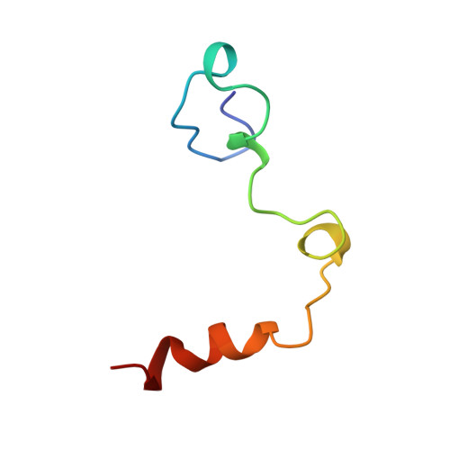
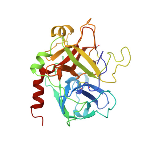
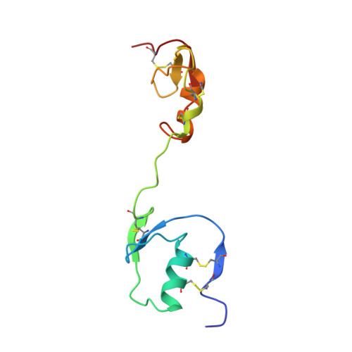
    Rhodniin is a highly specific inhibitor of thrombin isolated from the assassin bug Rhodnius prolixus. The 2.6 Angstrum crystal structure of the non-covalent complex between recombinant rhodniin and bovine alpha-thrombin reveals that the two Kazal-type domains of rhodniin bind to different sites of thrombin. The amino-terminal domain binds in a substrate-like manner to the narrow active-site cleft of thrombin; the imidazole group of the P1 His residue extends into the S1 pocket to form favourable hydrogen/ionic bonds with Asp189 at its bottom, and additionally with Glu192 at its entrance. The carboxy-terminal domain, whose distorted reactive-site loop cannot adopt the canonical conformation, docks to the fibrinogen recognition exosite via extensive electrostatic interactions. The rather acidic polypeptide linking the two domains is displaced from the thrombin surface, with none of its residues involved in direct salt bridges with thrombin. The tight (Ki = 2 x 10(-13) M) binding of rhodniin to thrombin is the result of the sum of steric and charge complementarity of the amino-terminal domain towards the active-site cleft, and of the electrostatic interactions between the carboxy-terminal domain and the exosite.


  • Organizational Affiliation

    Max-Planck-Institut für Biochemie, Abteilung Strukturforschung, Martinsried, Germany.


Macromolecules
Find similar proteins by:  (by identity cutoff)  |  3D Structure
Entity ID: 1
MoleculeChains Sequence LengthOrganismDetailsImage
THROMBINA [auth L],
C [auth J]
49Bos taurusMutation(s): 0 
EC: 3.4.21.5
UniProt
Find proteins for P00735 (Bos taurus)
Explore P00735 
Go to UniProtKB:  P00735
Entity Groups  
Sequence Clusters30% Identity50% Identity70% Identity90% Identity95% Identity100% Identity
UniProt GroupP00735
Sequence Annotations
Expand
  • Reference Sequence
Find similar proteins by:  (by identity cutoff)  |  3D Structure
Entity ID: 2
MoleculeChains Sequence LengthOrganismDetailsImage
THROMBINB [auth H],
D [auth K]
259Bos taurusMutation(s): 0 
EC: 3.4.21.5
UniProt
Find proteins for P00735 (Bos taurus)
Explore P00735 
Go to UniProtKB:  P00735
Entity Groups  
Sequence Clusters30% Identity50% Identity70% Identity90% Identity95% Identity100% Identity
UniProt GroupP00735
Sequence Annotations
Expand
  • Reference Sequence
Find similar proteins by:  (by identity cutoff)  |  3D Structure
Entity ID: 3
MoleculeChains Sequence LengthOrganismDetailsImage
RHODNIINE [auth R],
F [auth S]
103Rhodnius prolixusMutation(s): 0 
Gene Names: PRPTI
UniProt
Find proteins for Q06684 (Rhodnius prolixus)
Explore Q06684 
Go to UniProtKB:  Q06684
Entity Groups  
Sequence Clusters30% Identity50% Identity70% Identity90% Identity95% Identity100% Identity
UniProt GroupQ06684
Sequence Annotations
Expand
  • Reference Sequence
Experimental Data & Validation

Experimental Data

  • Method: X-RAY DIFFRACTION
  • Resolution: 3.10 Å
  • R-Value Work: 0.175 
  • R-Value Observed: 0.175 
  • Space Group: P 21 21 21
Unit Cell:
Length ( Å )Angle ( ˚ )
a = 91.311α = 90
b = 111.6β = 90
c = 112.14γ = 90
Software Package:
Software NamePurpose
MOSFLMdata reduction
X-PLORmodel building
X-PLORrefinement
X-PLORphasing

Structure Validation

View Full Validation Report


Entry History 

Deposition Data

Revision History  (Full details and data files)

  • Version 1.0: 1996-10-14
    Type: Initial release
  • Version 1.1: 2008-03-24
    Changes: Version format compliance
  • Version 1.2: 2011-07-13
    Changes: Version format compliance