1TA3

Crystal Structure of xylanase (GH10) in complex with inhibitor (XIP)


Experimental Data Snapshot

  • Method: X-RAY DIFFRACTION
  • Resolution: 1.70 Å
  • R-Value Free: 0.166 
  • R-Value Work: 0.133 
  • R-Value Observed: 0.135 

wwPDB Validation   3D Report Full Report


Ligand Structure Quality Assessment 


This is version 1.3 of the entry. See complete history


Literature

The Dual Nature of the Wheat Xylanase Protein Inhibitor XIP-I: STRUCTURAL BASIS FOR THE INHIBITION OF FAMILY 10 AND FAMILY 11 XYLANASES.

Payan, F.Leone, P.Porciero, S.Furniss, C.Tahir, T.Williamson, G.Durand, A.Manzanares, P.Gilbert, H.J.Juge, N.Roussel, A.

(2004) J Biol Chem 279: 36029-36037

  • DOI: https://doi.org/10.1074/jbc.M404225200
  • Primary Citation of Related Structures:  
    1TA3, 1TE1

  • PubMed Abstract: 

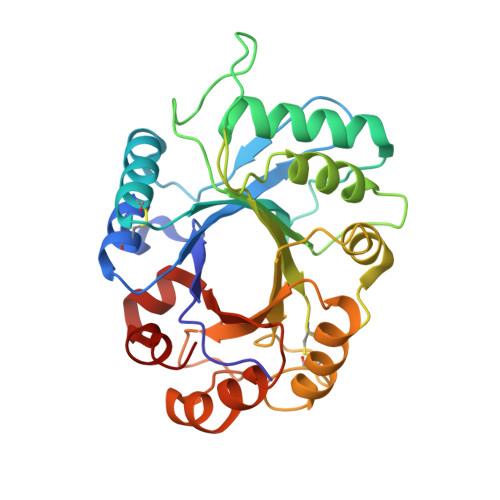
    The xylanase inhibitor protein I (XIP-I) from wheat Triticum aestivum is the prototype of a novel class of cereal protein inhibitors that inhibit fungal xylanases belonging to glycoside hydrolase families 10 (GH10) and 11 (GH11). The crystal structures of XIP-I in complex with Aspergillus nidulans (GH10) and Penicillium funiculosum (GH11) xylanases have been solved at 1.7 and 2.5 A resolution, respectively. The inhibition strategy is novel because XIP-I possesses two independent enzyme-binding sites, allowing binding to two glycoside hydrolases that display a different fold. Inhibition of the GH11 xylanase is mediated by the insertion of an XIP-I Pi-shaped loop (Lalpha(4)beta(5)) into the enzyme active site, whereas residues in the helix alpha7 of XIP-I, pointing into the four central active site subsites, are mainly responsible for the reversible inactivation of GH10 xylanases. The XIP-I strategy for inhibition of xylanases involves substrate-mimetic contacts and interactions occluding the active site. The structural determinants of XIP-I specificity demonstrate that the inhibitor is able to interact with GH10 and GH11 xylanases of both fungal and bacterial origin. The biological role of the xylanase inhibitors is discussed in light of the present structural data.


  • Organizational Affiliation

    Architecture et Fonction de Macromolécules Biologiques, UMR-6098, CNRS et Universités d'Aix-Marseille I et II, 31 Chemin Joseph Aiguier, 13402 Marseille Cedex 20, France.


Macromolecules
Find similar proteins by:  (by identity cutoff)  |  3D Structure
Entity ID: 1
MoleculeChains Sequence LengthOrganismDetailsImage
xylanase inhibitor protein I274Triticum aestivumMutation(s): 0 
UniProt
Find proteins for Q8L5C6 (Triticum aestivum)
Explore Q8L5C6 
Go to UniProtKB:  Q8L5C6
Entity Groups  
Sequence Clusters30% Identity50% Identity70% Identity90% Identity95% Identity100% Identity
UniProt GroupQ8L5C6
Sequence Annotations
Expand
  • Reference Sequence
Find similar proteins by:  (by identity cutoff)  |  3D Structure
Entity ID: 2
MoleculeChains Sequence LengthOrganismDetailsImage
Endo-1,4-beta-xylanase303Aspergillus nidulansMutation(s): 0 
Gene Names: XLNC
EC: 3.2.1.8
UniProt
Find proteins for Q00177 (Emericella nidulans (strain FGSC A4 / ATCC 38163 / CBS 112.46 / NRRL 194 / M139))
Explore Q00177 
Go to UniProtKB:  Q00177
Entity Groups  
Sequence Clusters30% Identity50% Identity70% Identity90% Identity95% Identity100% Identity
UniProt GroupQ00177
Sequence Annotations
Expand
  • Reference Sequence
Small Molecules
Ligands 2 Unique
IDChains Name / Formula / InChI Key2D Diagram3D Interactions
NAG
Query on NAG

Download Ideal Coordinates CCD File 
C [auth A],
D [auth A]
2-acetamido-2-deoxy-beta-D-glucopyranose
C8 H15 N O6
OVRNDRQMDRJTHS-FMDGEEDCSA-N
EDO
Query on EDO

Download Ideal Coordinates CCD File 
E [auth A]
F [auth B]
G [auth B]
H [auth B]
I [auth B]
E [auth A],
F [auth B],
G [auth B],
H [auth B],
I [auth B],
J [auth B],
K [auth B]
1,2-ETHANEDIOL
C2 H6 O2
LYCAIKOWRPUZTN-UHFFFAOYSA-N
Experimental Data & Validation

Experimental Data

  • Method: X-RAY DIFFRACTION
  • Resolution: 1.70 Å
  • R-Value Free: 0.166 
  • R-Value Work: 0.133 
  • R-Value Observed: 0.135 
  • Space Group: P 21 21 21
Unit Cell:
Length ( Å )Angle ( ˚ )
a = 46.73α = 90
b = 75.872β = 90
c = 159.654γ = 90
Software Package:
Software NamePurpose
REFMACrefinement
DENZOdata reduction
SCALEPACKdata scaling
AMoREphasing

Structure Validation

View Full Validation Report


Ligand Structure Quality Assessment 


Entry History 

Deposition Data

Revision History  (Full details and data files)

  • Version 1.0: 2004-07-20
    Type: Initial release
  • Version 1.1: 2008-04-30
    Changes: Version format compliance
  • Version 1.2: 2011-07-13
    Changes: Non-polymer description, Version format compliance
  • Version 1.3: 2020-07-29
    Type: Remediation
    Reason: Carbohydrate remediation
    Changes: Data collection, Derived calculations, Structure summary