1T8T

Crystal Structure of human 3-O-Sulfotransferase-3 with bound PAP


Experimental Data Snapshot

  • Method: X-RAY DIFFRACTION
  • Resolution: 1.85 Å
  • R-Value Free: 0.236 
  • R-Value Work: 0.206 
  • R-Value Observed: 0.206 

wwPDB Validation   3D Report Full Report


Ligand Structure Quality Assessment 


This is version 1.4 of the entry. See complete history


Literature

Structural analysis of the sulfotransferase (3-o-sulfotransferase isoform 3) involved in the biosynthesis of an entry receptor for herpes simplex virus 1

Moon, A.F.Edavettal, S.C.Krahn, J.M.Munoz, E.M.Negishi, M.Linhardt, R.J.Liu, J.Pedersen, L.C.

(2004) J Biol Chem 279: 45185-45193

  • DOI: https://doi.org/10.1074/jbc.M405013200
  • Primary Citation of Related Structures:  
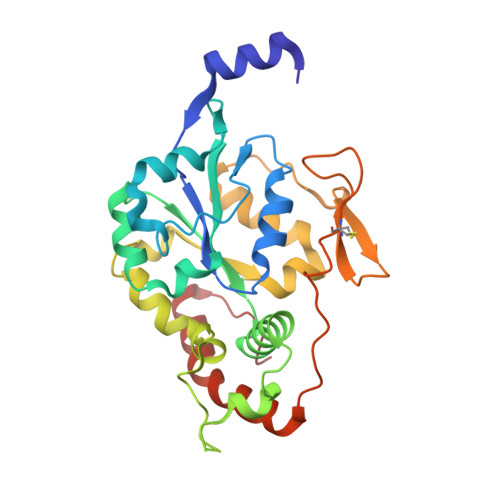
    1T8T

  • PubMed Abstract: 

    Heparan sulfate (HS) plays essential roles in assisting herpes simplex virus infection and other biological processes. The biosynthesis of HS includes numerous specialized sulfotransferases that generate a variety of sulfated saccharide sequences, conferring the selectivity of biological functions of HS. We report a structural study of human HS 3-O-sulfotransferase isoform 3 (3-OST-3), a key sulfotransferase that transfers a sulfuryl group to a specific glucosamine in HS generating an entry receptor for herpes simplex virus 1. We have obtained the crystal structure of 3-OST-3 at 1.95 A in a ternary complex with 3'-phosphoadenosine 5'-phosphate and a tetrasaccharide substrate. Mutational analyses were also performed on the residues involved in the binding of the substrate. Residues Gln255 and Lys368 are essential for the sulfotransferase activity and lie within hydrogen bonding distances to the carboxyl and sulfo groups of the uronic acid unit. These residues participate in the substrate recognition of 3-OST-3. This structure provides atomic level evidence for delineating the substrate recognition and catalytic mechanism for 3-OST-3.


  • Organizational Affiliation

    Laboratories of Structural Biology, NIEHS, National Institutes of Health, Research Triangle Park, North Carolina 27709, USA.


Macromolecules
Find similar proteins by:  (by identity cutoff)  |  3D Structure
Entity ID: 1
MoleculeChains Sequence LengthOrganismDetailsImage
heparan sulfate D-glucosaminyl 3-O-sulfotransferase 3A1
A, B
271Homo sapiensMutation(s): 0 
Gene Names: h3-OST-3
UniProt & NIH Common Fund Data Resources
Find proteins for Q9Y663 (Homo sapiens)
Explore Q9Y663 
Go to UniProtKB:  Q9Y663
PHAROS:  Q9Y663
GTEx:  ENSG00000153976 
Entity Groups  
Sequence Clusters30% Identity50% Identity70% Identity90% Identity95% Identity100% Identity
UniProt GroupQ9Y663
Sequence Annotations
Expand
  • Reference Sequence
Experimental Data & Validation

Experimental Data

  • Method: X-RAY DIFFRACTION
  • Resolution: 1.85 Å
  • R-Value Free: 0.236 
  • R-Value Work: 0.206 
  • R-Value Observed: 0.206 
  • Space Group: C 2 2 21
Unit Cell:
Length ( Å )Angle ( ˚ )
a = 80.338α = 90
b = 155.394β = 90
c = 93.323γ = 90
Software Package:
Software NamePurpose
CNSrefinement
HKL-2000data scaling
MOLREPphasing

Structure Validation

View Full Validation Report


Ligand Structure Quality Assessment 


Entry History 

Deposition Data

Revision History  (Full details and data files)

  • Version 1.0: 2004-08-31
    Type: Initial release
  • Version 1.1: 2008-04-30
    Changes: Version format compliance
  • Version 1.2: 2011-07-13
    Changes: Version format compliance
  • Version 1.3: 2017-10-11
    Changes: Refinement description
  • Version 1.4: 2023-08-23
    Changes: Data collection, Database references, Derived calculations, Refinement description