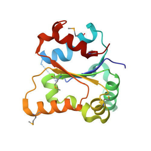
Crystal structure of the Toll/interleukin-1 receptor domain of human IL-1RAPL
Khan, J.A., Brint, E.K., O'Neill, L.A.J., Tong, L.(2004) J Biol Chem 279: 31664-31670
- PubMed: 15123616
- DOI: https://doi.org/10.1074/jbc.M403434200
- Primary Citation of Related Structures:
1T3G - PubMed Abstract:
The Toll/interleukin-1 receptor (TIR) domain is conserved in the intracellular regions of Toll-like receptors (TLRs) and interleukin-1 receptors (IL-1Rs) as well as in several cytoplasmic adapter molecules. This domain has crucial roles in signal transduction by these receptors for host immune response. Here we report the crystal structure at 2.3-A resolution of the TIR domain of human IL-1RAPL, the first structure of a TIR domain of the IL-1R superfamily. There are large structural differences between this TIR domain and that of TLR1 and TLR2. Helix alphaD in IL-1RAPL is almost perpendicular to its equivalent in TLR1 or TLR2. The BB loop contains a hydrogen bond unique to IL-1RAPL between Thr residues at the 8th and 10th positions. The structural and sequence diversity among these domains may be important for specificity in the signal transduction by these receptors. A dimer of the TIR domain of IL-1RAPL is observed in the crystal, although this domain is monomeric in solution. Residues in the dimer interface are mostly unique to IL-1RAPL, which is consistent with the distinct functional roles of this receptor. Our functional studies show IL-1RAPL can activate JNK but not the ERK or the p38 MAP kinases, whereas its close homolog, TIGIRR, cannot activate JNK. Deletion mutagenesis studies show that the activation of JNK by IL-1RAPL does not depend on the integrity of its TIR domain, suggesting a distinct mechanism of signaling through this receptor.
Organizational Affiliation:
Department of Biological Sciences, Columbia University, New York, New York 10027, USA