1T1V

Crystal Structure of the Glutaredoxin-like Protein SH3BGRL3 at 1.6 A resolution


Experimental Data Snapshot

  • Method: X-RAY DIFFRACTION
  • Resolution: 1.60 Å
  • R-Value Free: 0.244 
  • R-Value Work: 0.197 
  • R-Value Observed: 0.202 

wwPDB Validation   3D Report Full Report


This is version 1.3 of the entry. See complete history


Literature

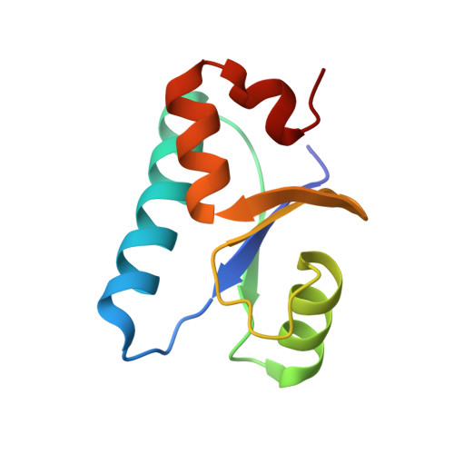
Crystal structure of the glutaredoxin-like protein SH3BGRL3 at 1.6 A resolution

Nardini, M.Mazzocco, M.Massaro, M.Maffei, M.Vergano, A.Donadini, A.Scartezzini, M.Bolognesi, M.

(2004) Biochem Biophys Res Commun 318: 470-476

  • DOI: https://doi.org/10.1016/j.bbrc.2004.04.050
  • Primary Citation of Related Structures:  
    1T1V

  • PubMed Abstract: 

    We report the 1.6 Angstrom resolution crystal structure of SH3BGRL3, a member of a new mammalian protein family of unknown function. The observed "thioredoxin fold" of SH3BGRL3 matches the tertiary structure of glutaredoxins, even in the N-terminal region where the sequence similarity between the two protein families is negligible. In particular, SH3BGRL3 displays structural modifications at the N-terminal Cys-x-x-Cys loop, responsible for glutathione binding and catalysis in glutaredoxins. The loop hosts a six residue insertion, yielding an extra N-terminal-capped helical turn, first observed here for the thioredoxin fold. This, together with deletion of both Cys residues, results in a substantial reshaping of the neighboring cleft, where glutathione is hosted in glutaredoxins. While not active in redox reaction and glutathione binding, SH3BGRL3 may act as an endogenous modulator of glutaredoxin activities by competing, with its fully conserved thioredoxin fold, for binding to yet unknown target proteins.


  • Organizational Affiliation

    Dipartimento di Fisica-INFM e Centro di Eccellenza per la Ricerca Biomedica, Università di Genova, Via Dodecaneso 33, 16146 Genoa, Italy.


Macromolecules
Find similar proteins by:  (by identity cutoff)  |  3D Structure
Entity ID: 1
MoleculeChains Sequence LengthOrganismDetailsImage
SH3 domain-binding glutamic acid-rich protein-like 3
A, B
93Mus musculusMutation(s): 0 
UniProt & NIH Common Fund Data Resources
Find proteins for Q91VW3 (Mus musculus)
Explore Q91VW3 
Go to UniProtKB:  Q91VW3
IMPC:  MGI:1920973
Entity Groups  
Sequence Clusters30% Identity50% Identity70% Identity90% Identity95% Identity100% Identity
UniProt GroupQ91VW3
Sequence Annotations
Expand
  • Reference Sequence
Experimental Data & Validation

Experimental Data

  • Method: X-RAY DIFFRACTION
  • Resolution: 1.60 Å
  • R-Value Free: 0.244 
  • R-Value Work: 0.197 
  • R-Value Observed: 0.202 
  • Space Group: P 1 21 1
Unit Cell:
Length ( Å )Angle ( ˚ )
a = 29.624α = 90
b = 80.38β = 93.96
c = 34.66γ = 90
Software Package:
Software NamePurpose
DENZOdata reduction
SCALEPACKdata scaling
MOLREPphasing
REFMACrefinement

Structure Validation

View Full Validation Report


Entry History 

Deposition Data

Revision History  (Full details and data files)

  • Version 1.0: 2004-06-29
    Type: Initial release
  • Version 1.1: 2008-04-30
    Changes: Version format compliance
  • Version 1.2: 2011-07-13
    Changes: Non-polymer description, Version format compliance
  • Version 1.3: 2023-08-23
    Changes: Data collection, Database references, Derived calculations, Refinement description