Multiple active site conformations revealed by distant site mutation in ornithine decarboxylase
Jackson, L.K., Baldwin, J., Akella, R., Goldsmith, E.J., Phillips, M.A.(2004) Biochemistry 43: 12990-12999
- PubMed: 15476392
- DOI: https://doi.org/10.1021/bi048933l
- Primary Citation of Related Structures:
1SZR - PubMed Abstract:
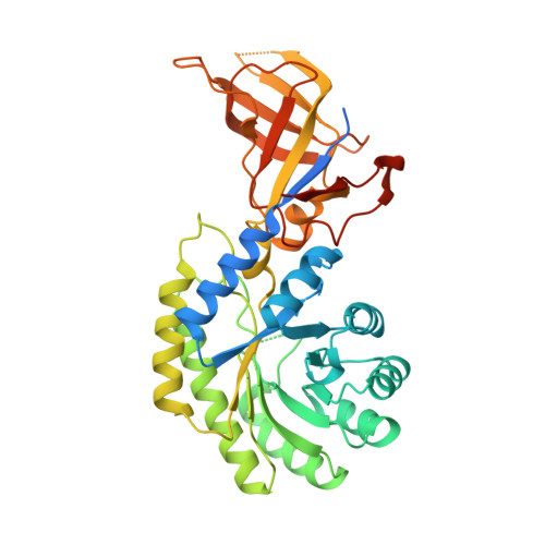
Ornithine decarboxylase (ODC) is an obligate homodimer that catalyzes the pyridoxal 5'-phosphate-dependent decarboxylation of l-ornithine to putrescine, a vital step in polyamine biosynthesis. A previous mutagenic analysis of the ODC dimer interface identified several residues that were distant from the active site yet had a greater impact on catalytic activity than on dimer stability [Myers, D. P., et al. (2001) Biochemistry 40, 13230-13236]. To better understand the basis of this phenomenon, the structure of the Trypanosoma brucei ODC mutant K294A was determined to 2.15 A resolution in complex with the substrate analogue d-ornithine. This residue is distant from the reactive center (>10 A from the PLP Schiff base), and its mutation reduced catalytic efficiency by 3 kcal/mol. The X-ray structure demonstrates that the mutation increases the disorder of residues Leu-166-Ala-172 (Lys-169 loop), which normally form interactions with Lys-294 across the dimer interface. In turn, the Lys-169 loop forms interactions with the active site, suggesting that the reduced catalytic efficiency is mediated by the decreased stability of this loop. The extent of disorder varies in the four Lys-169 loops in the asymmetric unit, suggesting that the mutation has led to an increase in the population of inactive conformations. The structure also reveals that the mutation has affected the nature of the ligand-bound species. Each of the four active sites contains unusual ligands. The electron density suggests one active site contains a gem-diamine intermediate with d-ornithine; the second has density consistent with a tetrahedral adduct with glycine, and the remaining two contain tetrahedral adducts of PLP, Lys-69, and water (or hydroxide). These data also suggest that the structure is less constrained in the mutant enzyme. The observation of a gem-diamine intermediate provides insight into the conformational changes that occur during the ODC catalytic cycle.
Organizational Affiliation:
Department of Pharmacology, The University of Texas Southwestern Medical Center at Dallas, 5323 Harry Hines Boulevard, Dallas, Texas 75390-9041, USA.