1SZO

Crystal Structure Analysis of the 6-Oxo Camphor Hydrolase His122Ala Mutant Bound to Its Natural Product (2S,4S)-alpha-Campholinic Acid


Experimental Data Snapshot

  • Method: X-RAY DIFFRACTION
  • Resolution: 1.90 Å
  • R-Value Free: 0.198 
  • R-Value Work: 0.164 
  • R-Value Observed: 0.164 

wwPDB Validation   3D Report Full Report


Ligand Structure Quality Assessment 


This is version 1.4 of the entry. See complete history


Literature

Structure of 6-oxo camphor hydrolase H122A mutant bound to its natural product, (2S,4S)-alpha-campholinic acid: mutant structure suggests an atypical mode of transition state binding for a crotonase homolog.

Leonard, P.M.Grogan, G.

(2004) J Biol Chem 279: 31312-31317

  • DOI: https://doi.org/10.1074/jbc.M403514200
  • Primary Citation of Related Structures:  
    1SZO

  • PubMed Abstract: 

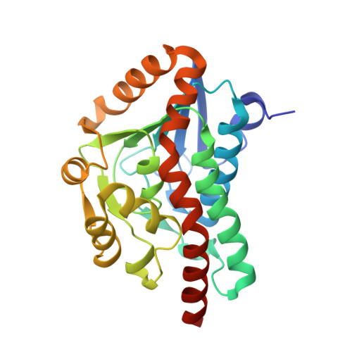
    The crotonase homolog, 6-oxo camphor hydrolase (OCH), catalyzes the desymmetrization of bicyclic beta-diketones to optically active keto acids via an enzymatic retro-Claisen reaction, resulting in the cleavage of a carbon-carbon bond. We have previously reported the structure of OCH (Whittingham, J. L., Turkenburg, J. P., Verma, C. S., Walsh, M. A., and Grogan, G. (2003) J. Biol. Chem. 278, 1744-1750), which suggested the involvement of five residues, His-45, His-122, His-145, Asp-154, and Glu-244, in catalysis. Here we report mutation studies on OCH that reveal that H145A and D154N mutants of OCH have greatly reduced values of k(cat)/K(m) derived from a very large increase in K(m) for the native substrate, 6-oxo camphor. In addition, H122A has a greatly reduced value of k(cat), and its K(m) is five times that of the wild-type. The location of the active site is confirmed by the 1.9-A structure of the H122A mutant of OCH complexed with the minor diastereoisomer of (2S,4S)-alpha-campholinic acid, the natural product of the enzyme. This shows the pendant acetate of the product hydrogen bonded to a His-145/Asp-154 dyad and the endocyclic carbonyl of the cyclopentane ring hydrogen bonded to Trp-40. The results are suggestive of a base-catalyzed mechanism of C-C bond cleavage and provide clues to the origin of prochiral selectivity by the enzyme and to the recruitment of the crotonase fold for alternate modes of transition state stabilization to those described for other crotonase superfamily members.


  • Organizational Affiliation

    York Structural Biology Laboratory, Department of Chemistry, University of York, Heslington, York YO10 5YW, United Kingdom.


Macromolecules
Find similar proteins by:  (by identity cutoff)  |  3D Structure
Entity ID: 1
MoleculeChains Sequence LengthOrganismDetailsImage
6-oxocamphor hydrolase
A, B, C, D, E
A, B, C, D, E, F, G, H, I, J, K, L
257Rhodococcus sp. NCIMB 9784Mutation(s): 1 
Gene Names: camK
UniProt
Find proteins for Q93TU6 (Rhodococcus sp)
Explore Q93TU6 
Go to UniProtKB:  Q93TU6
Entity Groups  
Sequence Clusters30% Identity50% Identity70% Identity90% Identity95% Identity100% Identity
UniProt GroupQ93TU6
Sequence Annotations
Expand
  • Reference Sequence
Small Molecules
Ligands 2 Unique
IDChains Name / Formula / InChI Key2D Diagram3D Interactions
CAX
Query on CAX

Download Ideal Coordinates CCD File 
AA [auth K]
BA [auth L]
M [auth A]
O [auth B]
P [auth C]
AA [auth K],
BA [auth L],
M [auth A],
O [auth B],
P [auth C],
Q [auth D],
R [auth E],
T [auth F],
U [auth G],
V [auth H],
X [auth I],
Y [auth J]
(2S,4S)-4-(2,2-DIHYDROXYETHYL)-2,3,3-TRIMETHYLCYCLOPENTANONE
C10 H18 O3
KAXFPJKKGITBPU-RQJHMYQMSA-N
CA
Query on CA

Download Ideal Coordinates CCD File 
N [auth B],
S [auth F],
W [auth I],
Z [auth K]
CALCIUM ION
Ca
BHPQYMZQTOCNFJ-UHFFFAOYSA-N
Experimental Data & Validation

Experimental Data

  • Method: X-RAY DIFFRACTION
  • Resolution: 1.90 Å
  • R-Value Free: 0.198 
  • R-Value Work: 0.164 
  • R-Value Observed: 0.164 
  • Space Group: P 1 21 1
Unit Cell:
Length ( Å )Angle ( ˚ )
a = 83.28α = 90
b = 132.008β = 94.11
c = 135.424γ = 90
Software Package:
Software NamePurpose
DENZOdata reduction
SCALEPACKdata scaling
AMoREphasing
REFMACrefinement

Structure Validation

View Full Validation Report


Ligand Structure Quality Assessment 


Entry History 

Deposition Data

Revision History  (Full details and data files)

  • Version 1.0: 2004-06-29
    Type: Initial release
  • Version 1.1: 2008-04-30
    Changes: Version format compliance
  • Version 1.2: 2011-07-13
    Changes: Version format compliance
  • Version 1.3: 2021-10-27
    Changes: Database references, Derived calculations
  • Version 1.4: 2023-08-23
    Changes: Data collection, Refinement description