1SZI

Crystal Structure of the C-terminus of TIP47


Experimental Data Snapshot

  • Method: X-RAY DIFFRACTION
  • Resolution: 2.80 Å
  • R-Value Free: 0.272 
  • R-Value Work: 0.236 
  • R-Value Observed: 0.238 

wwPDB Validation   3D Report Full Report


This is version 1.4 of the entry. See complete history


Literature

Structure of a Lipid Droplet Protein: The PAT Family Member TIP47

Hickenbottom, S.J.Kimmel, A.R.Londos, C.Hurley, J.H.

(2004) Structure 12: 1199-1207

  • DOI: https://doi.org/10.1016/j.str.2004.04.021
  • Primary Citation of Related Structures:  
    1SZI

  • PubMed Abstract: 

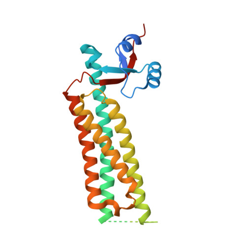
    The perilipin/ADRP/TIP47 (PAT) proteins localize to the surface of intracellular neutral lipid droplets. Perilipin is essential for lipid storage and hormone regulated lipolysis in adipocytes, and perilipin null mice exhibit a dramatic reduction in adipocyte lipid stores. A significant fraction of the approximately 200 amino acid N-terminal region of the PAT proteins consists of 11-mer helical repeats that are also found in apolipoproteins and other lipid-associated proteins. The C-terminal 60% of TIP47, a representative PAT protein, comprises a monomeric and independently folded unit. The crystal structure of the C-terminal portion of TIP47 was determined and refined at 2.8 A resolution. The structure consists of an alpha/beta domain of novel topology and a four-helix bundle resembling the LDL receptor binding domain of apolipoprotein E. The structure suggests an analogy between PAT proteins and apolipoproteins in which helical repeats interact with lipid while the ordered C-terminal region is involved in protein:protein interactions.


  • Organizational Affiliation

    Laboratory of Cellular and Developmental Biology, U.S. Department of Health and Human Services, Bethesda, MD 20892, USA.


Macromolecules
Find similar proteins by:  (by identity cutoff)  |  3D Structure
Entity ID: 1
MoleculeChains Sequence LengthOrganismDetailsImage
mannose-6-phosphate receptor binding protein 1247Mus musculusMutation(s): 0 
Membrane Entity: Yes 
UniProt
Find proteins for Q9DBG5 (Mus musculus)
Explore Q9DBG5 
Go to UniProtKB:  Q9DBG5
Entity Groups  
Sequence Clusters30% Identity50% Identity70% Identity90% Identity95% Identity100% Identity
UniProt GroupQ9DBG5
Sequence Annotations
Expand
  • Reference Sequence
Experimental Data & Validation

Experimental Data

  • Method: X-RAY DIFFRACTION
  • Resolution: 2.80 Å
  • R-Value Free: 0.272 
  • R-Value Work: 0.236 
  • R-Value Observed: 0.238 
  • Space Group: P 63 2 2
Unit Cell:
Length ( Å )Angle ( ˚ )
a = 118.746α = 90
b = 118.746β = 90
c = 97.291γ = 120
Software Package:
Software NamePurpose
REFMACrefinement
HKL-2000data scaling
SOLVEphasing
RESOLVEphasing
Omodel building

Structure Validation

View Full Validation Report


Entry History 

Deposition Data

Revision History  (Full details and data files)

  • Version 1.0: 2004-07-27
    Type: Initial release
  • Version 1.1: 2008-04-30
    Changes: Version format compliance
  • Version 1.2: 2011-07-13
    Changes: Version format compliance
  • Version 1.3: 2017-10-11
    Changes: Refinement description
  • Version 1.4: 2024-02-14
    Changes: Data collection, Database references