1SXQ

BGT in complex with a 13mer DNA containing a central C:G base pair and UDP


Experimental Data Snapshot

  • Method: X-RAY DIFFRACTION
  • Resolution: 1.80 Å
  • R-Value Free: 0.212 
  • R-Value Work: 0.183 

wwPDB Validation   3D Report Full Report


Ligand Structure Quality Assessment 


This is version 1.3 of the entry. See complete history


Literature

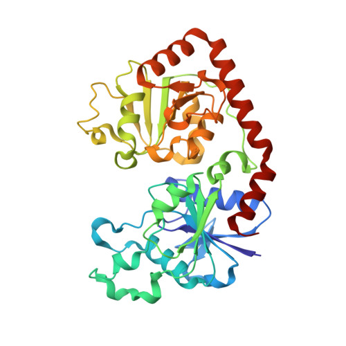
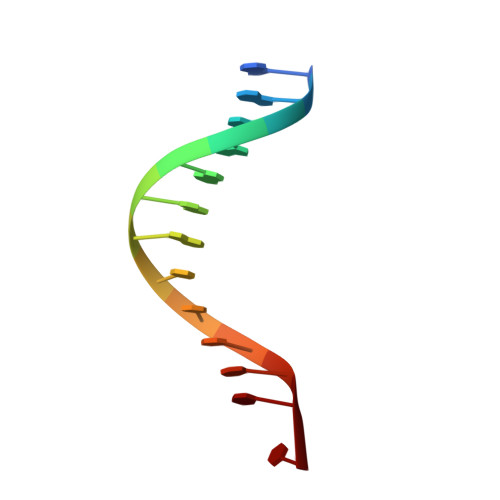
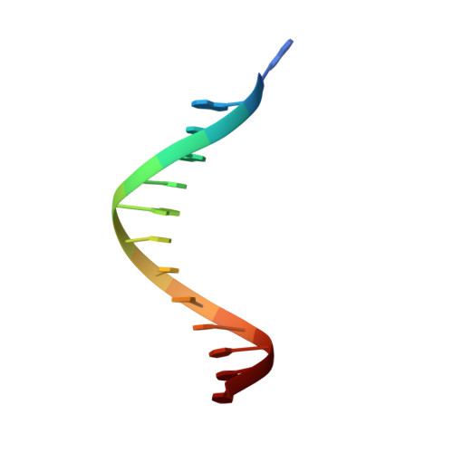
Structural evidence of a passive base flipping mechanism for {beta}-Glucosyltransferase

Lariviere, L.Morera, S.

(2004) J Biol Chem 279: 34715-34720

  • DOI: https://doi.org/10.1074/jbc.M404394200
  • Primary Citation of Related Structures:  
    1SXP, 1SXQ

  • PubMed Abstract: 

    Beta-glucosyltransferase (BGT) is a DNA-modifying enzyme and a glycosyltransferase. This inverting enzyme transfers glucose from UDP-glucose to the 5-hydroxymethyl cytosine bases of T4 phage DNA. From previous structural analyses we showed that Asp-100 and Asn-70 were, respectively, the catalytic base and the key residue for specific DNA recognition (Larivière, L., Gueguen-Chaignon, V., and Moréra, S. (2003) J. Mol. Biol. 330, 1077-1086). Here, we supply biochemical evidence supporting their essential roles in catalysis. We have also shown previously that BGT uses a base-flipping mechanism to access 5-hydroxymethyl cytosine (Larivière, L., and Moréra, S. (2002) J. Mol. Biol. 324, 483-490). Whether it is an active or a passive process remains unclear, as is the case for all DNA cleaving and modifying enzymes. Here, we report two crystal structures: (i) BGT in complex with a 13-mer DNA containing an A:G mismatch and (ii) BGT in a ternary complex with UDP and an oligonucleotide containing a single central G:C base pair. The binary structure reveals a specific complex with the flipped-out, mismatched adenine exposed to the active site. Unexpectedly, the other structure shows the non-productive binding of an intermediate flipped-out base. Our structural analysis provides clear evidence for a passive process.


  • Organizational Affiliation

    Laboratoire d'Enzymologie et Biochimie Structurales, Unité Propre de Recherche 9063 CNRS, Bātiment 34, 1 Avenue de la Terrasse, 91198 Gif-sur-Yvette, France.


Macromolecules

Find similar proteins by:  (by identity cutoff)  |  3D Structure
Entity ID: 3
MoleculeChains Sequence LengthOrganismDetailsImage
DNA beta-glucosyltransferaseE [auth A],
F [auth B]
351Tequatrovirus T4Mutation(s): 0 
Gene Names: BGTBETA-GT
EC: 2.4.1.27
UniProt
Find proteins for P04547 (Enterobacteria phage T4)
Explore P04547 
Go to UniProtKB:  P04547
Entity Groups  
Sequence Clusters30% Identity50% Identity70% Identity90% Identity95% Identity100% Identity
UniProt GroupP04547
Sequence Annotations
Expand
  • Reference Sequence

Find similar nucleic acids by:  Sequence   |   3D Structure  

Entity ID: 1
MoleculeChains LengthOrganismImage
5'-D(*AP*AP*AP*AP*AP*AP*GP*TP*TP*TP*TP*TP*T)-3'A [auth C],
C [auth D]
13N/A
Sequence Annotations
Expand
  • Reference Sequence

Find similar nucleic acids by:  Sequence   |   3D Structure  

Entity ID: 2
MoleculeChains LengthOrganismImage
5'-D(*AP*AP*AP*AP*AP*AP*CP*TP*TP*TP*TP*TP*T)-3'B [auth E],
D [auth F]
13N/A
Sequence Annotations
Expand
  • Reference Sequence
Small Molecules
Experimental Data & Validation

Experimental Data

  • Method: X-RAY DIFFRACTION
  • Resolution: 1.80 Å
  • R-Value Free: 0.212 
  • R-Value Work: 0.183 
  • Space Group: P 1 21 1
Unit Cell:
Length ( Å )Angle ( ˚ )
a = 72.652α = 90
b = 70.157β = 90.55
c = 96.581γ = 90
Software Package:
Software NamePurpose
DENZOdata reduction
SCALEPACKdata scaling
AMoREphasing
CNSrefinement

Structure Validation

View Full Validation Report


Ligand Structure Quality Assessment 


Entry History 

Deposition Data

Revision History  (Full details and data files)

  • Version 1.0: 2004-06-22
    Type: Initial release
  • Version 1.1: 2008-04-30
    Changes: Version format compliance
  • Version 1.2: 2011-07-13
    Changes: Version format compliance
  • Version 1.3: 2024-02-14
    Changes: Data collection, Database references, Derived calculations