1ST8

Crystal structure of fructan 1-exohydrolase IIa from Cichorium intybus


Experimental Data Snapshot

  • Method: X-RAY DIFFRACTION
  • Resolution: 2.35 Å
  • R-Value Free: 0.200 
  • R-Value Work: 0.183 
  • R-Value Observed: 0.183 

wwPDB Validation   3D Report Full Report


This is version 2.0 of the entry. See complete history


Literature

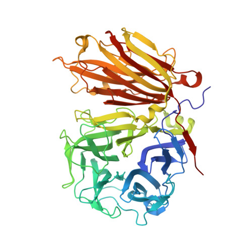
X-ray diffraction structure of a plant glycosyl hydrolase family 32 protein: fructan 1-exohydrolase IIa of Cichorium intybus.

Verhaest, M.van den Ende, W.Roy, K.L.De Ranter, C.J.van Laere, A.Rabijns, A.

(2005) Plant J 41: 400-411

  • DOI: https://doi.org/10.1111/j.1365-313X.2004.02304.x
  • Primary Citation of Related Structures:  
    1ST8

  • PubMed Abstract: 

    Fructan 1-exohydrolase, an enzyme involved in fructan degradation, belongs to the glycosyl hydrolase family 32. The structure of isoenzyme 1-FEH IIa from Cichorium intybus is described at a resolution of 2.35 A. The structure consists of an N-terminal fivefold beta-propeller domain connected to two C-terminal beta-sheets. The putative active site is located entirely in the beta-propeller domain and is formed by amino acids which are highly conserved within glycosyl hydrolase family 32. The fructan-binding site is thought to be in the cleft formed between the two domains. The 1-FEH IIa structure is compared with the structures of two homologous but functionally different enzymes: a levansucrase from Bacillus subtilis (glycosyl hydrolase family 68) and an invertase from Thermotoga maritima (glycosyl hydrolase family 32).


  • Organizational Affiliation

    Laboratorium voor Analytische Chemie en Medicinale Fysicochemie, Faculteit Farmaceutische Wetenschappen, K.U. Leuven, E. Van Evenstraat 4, B-3000 Leuven, Belgium.


Macromolecules
Find similar proteins by:  (by identity cutoff)  |  3D Structure
Entity ID: 1
MoleculeChains Sequence LengthOrganismDetailsImage
fructan 1-exohydrolase IIa543Cichorium intybusMutation(s): 0 
Gene Names: 1-feh IIa
EC: 3.2.1.80
UniProt
Find proteins for Q93X60 (Cichorium intybus)
Explore Q93X60 
Go to UniProtKB:  Q93X60
Entity Groups  
Sequence Clusters30% Identity50% Identity70% Identity90% Identity95% Identity100% Identity
UniProt GroupQ93X60
Sequence Annotations
Expand
  • Reference Sequence
Oligosaccharides

Help

Entity ID: 2
MoleculeChains Length2D Diagram Glycosylation3D Interactions
alpha-D-mannopyranose-(1-4)-2-acetamido-2-deoxy-beta-D-glucopyranose-(1-4)-2-acetamido-2-deoxy-beta-D-glucopyranose
B
3N-Glycosylation
Glycosylation Resources
GlyTouCan:  G62182OO
GlyCosmos:  G62182OO
GlyGen:  G62182OO
Entity ID: 3
MoleculeChains Length2D Diagram Glycosylation3D Interactions
2-acetamido-2-deoxy-beta-D-glucopyranose-(1-4)-2-acetamido-2-deoxy-beta-D-glucopyranose
C
2N-Glycosylation
Glycosylation Resources
GlyTouCan:  G42666HT
GlyCosmos:  G42666HT
GlyGen:  G42666HT
Experimental Data & Validation

Experimental Data

  • Method: X-RAY DIFFRACTION
  • Resolution: 2.35 Å
  • R-Value Free: 0.200 
  • R-Value Work: 0.183 
  • R-Value Observed: 0.183 
  • Space Group: P 41 21 2
Unit Cell:
Length ( Å )Angle ( ˚ )
a = 139.83α = 90
b = 139.83β = 90
c = 181.94γ = 90
Software Package:
Software NamePurpose
CNSrefinement
DENZOdata reduction
SCALEPACKdata scaling
SHARPphasing

Structure Validation

View Full Validation Report


Entry History 

Deposition Data

Revision History  (Full details and data files)

  • Version 1.0: 2005-03-01
    Type: Initial release
  • Version 1.1: 2008-04-29
    Changes: Version format compliance
  • Version 1.2: 2011-07-13
    Changes: Non-polymer description, Version format compliance
  • Version 2.0: 2020-07-29
    Type: Remediation
    Reason: Carbohydrate remediation
    Changes: Advisory, Atomic model, Data collection, Derived calculations, Non-polymer description, Structure summary