Nuclear magnetic resonance solution structure of the plasminogen-activator protein staphylokinase.
Ohlenschlager, O., Ramachandran, R., Guhrs, K.H., Schlott, B., Brown, L.R.(1998) Biochemistry 37: 10635-10642
- PubMed: 9692953
- DOI: https://doi.org/10.1021/bi980673i
- Primary Citation of Related Structures:
1SSN - PubMed Abstract:
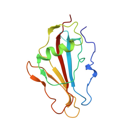
Staphylokinase, a 15.5 kDa protein from Staphylococcus aureus, is a plasminogen activator which is currently undergoing clinical trials for the therapy of myocardial infarction and peripheral thrombosis. The three-dimensional (3D) NMR solution structure has been determined by multidimensional heteronuclear NMR spectroscopy on uniformly 15N- and 15N,13C-labeled samples of staphylokinase. Structural constraints were obtained from 82 3JHNH alpha as well as 22 3JNH beta scalar coupling constants and 2345 NOE cross-peaks, derived from 15N-edited and 13C-edited 3D NOE spectra. NOE cross-peak assignments were confirmed by analysis of ¿15N,13C¿-edited and ¿13C,13C¿-edited 4D NOE spectra. The structure is presented as a family of 20 conformers which show an average rmsd of 1.02 +/- 0.15 A from the mean structure for the backbone atoms. The tertiary structure of staphylokinase shows a well-defined global structure consisting of a central 13-residue alpha-helix flanked by a two-stranded beta-sheet, both of which are located above a five-stranded beta-sheet. Two of the connecting loops exhibit a higher conformational heterogeneity. Overall, staphylokinase shows a strong asymmetry of hydrophilic and hydrophobic surfaces. The N-terminal sequence, including Lys10 which is the site of the initial proteolytic cleavage during activation of plasminogen, folds back onto the protein core, thereby shielding amino acids with functional importance in the plasminogen activation process. From a comparison of the structure with mutational studies, a binding region for plasminogen is proposed.
Organizational Affiliation:
Department of Molecular Biophysics and NMR Spectroscopy, Institute for Molecular Biotechnology, Jena, Germany.