1SRU

Crystal structure of full length E. coli SSB protein


Experimental Data Snapshot

  • Method: X-RAY DIFFRACTION
  • Resolution: 3.30 Å
  • R-Value Free: 0.309 
  • R-Value Work: 0.288 
  • R-Value Observed: 0.288 

wwPDB Validation   3D Report Full Report


This is version 1.3 of the entry. See complete history


Literature

The C-terminal domain of full-length E. coli SSB is disordered even when bound to DNA.

Savvides, S.N.Raghunathan, S.Fuetterer, K.Kozlov, A.G.Lohman, T.M.Waksman, G.

(2004) Protein Sci 13: 1942-1947

  • DOI: https://doi.org/10.1110/ps.04661904
  • Primary Citation of Related Structures:  
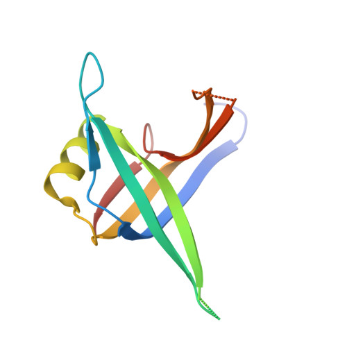
    1SRU

  • PubMed Abstract: 

    The crystal structure of full-length homotetrameric single-stranded DNA (ssDNA)-binding protein from Escherichia coli (SSB) has been determined to 3.3 A resolution and reveals that the entire C-terminal domain is disordered even in the presence of ssDNA. To our knowledge, this is the first experimental evidence that the C-terminal domain of SSB may be inherently disordered. The N-terminal DNA-binding domain of the protein is well ordered and is virtually indistinguishable from the previously determined structure of the chymotryptic fragment of SSB (SSBc) in complex with ssDNA. The absence of observable interactions with the core protein and the crystal packing of SSB together suggest that the disordered C-terminal domains likely extend laterally away from the DNA- binding domains, which may facilitate interactions with components of the replication machinery in vivo. The structure also reveals the conservation of molecular contacts between successive tetramers mediated by the L(45) loops as seen in two other crystal forms of SSBc, suggesting a possible functional relevance of this interaction.


  • Organizational Affiliation

    Department of Biochemistry and Molecular Biophysics, Washington University School of Medicine, St. Louis, MO 63110, USA.


Macromolecules
Find similar proteins by:  (by identity cutoff)  |  3D Structure
Entity ID: 1
MoleculeChains Sequence LengthOrganismDetailsImage
Single-strand binding protein
A, B, C, D
113Escherichia coliMutation(s): 0 
Gene Names: SSBEXRBLEXCB4059C5049Z5658ECS5041SF4145S3584
UniProt
Find proteins for P0AGE0 (Escherichia coli (strain K12))
Explore P0AGE0 
Go to UniProtKB:  P0AGE0
Entity Groups  
Sequence Clusters30% Identity50% Identity70% Identity90% Identity95% Identity100% Identity
UniProt GroupP0AGE0
Sequence Annotations
Expand
  • Reference Sequence
Experimental Data & Validation

Experimental Data

  • Method: X-RAY DIFFRACTION
  • Resolution: 3.30 Å
  • R-Value Free: 0.309 
  • R-Value Work: 0.288 
  • R-Value Observed: 0.288 
  • Space Group: P 31 1 2
Unit Cell:
Length ( Å )Angle ( ˚ )
a = 60.85α = 90
b = 60.85β = 90
c = 348.92γ = 120
Software Package:
Software NamePurpose
CNSrefinement
DENZOdata reduction
SCALEPACKdata scaling
CNSphasing

Structure Validation

View Full Validation Report


Entry History 

Deposition Data

Revision History  (Full details and data files)

  • Version 1.0: 2004-08-03
    Type: Initial release
  • Version 1.1: 2008-04-29
    Changes: Version format compliance
  • Version 1.2: 2011-07-13
    Changes: Version format compliance
  • Version 1.3: 2024-02-14
    Changes: Data collection, Database references