1SQJ

Crystal Structure Analysis of Oligoxyloglucan reducing-end-specific cellobiohydrolase (OXG-RCBH)


Experimental Data Snapshot

  • Method: X-RAY DIFFRACTION
  • Resolution: 2.20 Å
  • R-Value Free: 0.251 
  • R-Value Work: 0.217 
  • R-Value Observed: 0.219 

wwPDB Validation   3D Report Full Report


This is version 1.2 of the entry. See complete history


Literature

Tandem Repeat of a Seven-Bladed beta-Propeller Domain in Oligoxyloglucan Reducing-End-Specific Cellobiohydrolase

Yaoi, K.Kondo, H.Noro, N.Suzuki, M.Tsuda, S.Mitsuishi, Y.

(2004) Structure 12: 1209-1217

  • DOI: https://doi.org/10.1016/j.str.2004.04.020
  • Primary Citation of Related Structures:  
    1SQJ

  • PubMed Abstract: 

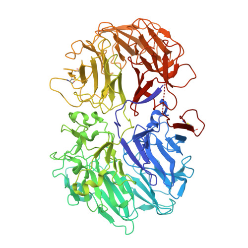
    Oligoxyloglucan reducing-end-specific cellobiohydrolase (OXG-RCBH; EC 3.2.1.150) is an exoglucanase that recognizes the reducing end of oligoxyloglucan and releases two glucosyl residue segments from the main chain. The X-ray crystal structure of OXG-RCBH determined at 2.2 A resolution reveals a unique feature of this enzyme; OXG-RCBH consists of a tandem repeat of two similar domains, which are both folded into seven-bladed beta-propeller structures. The sequence alignment of the propeller blades, based on the structure, indicates that a weak repeat of the amino acid sequence occurred seven times to construct each domain. There is a cleft that can accommodate the substrate oligosaccharide between the two domains, which is a putative substrate binding subsite. Mutation of either Asp35 or Asp465, located in the putative catalytic center, to Asn resulted in a protein with no detectable catalytic activity, indicating the critical role of these amino acids in catalysis.


  • Organizational Affiliation

    Institute for Biological Resources and Functions, National Institute of Advanced Industrial Science and Technology (AIST), Tsukuba Central 6, 1-1-1 Higashi, Tsukuba, Ibaraki 305-8566, Japan.


Macromolecules
Find similar proteins by:  (by identity cutoff)  |  3D Structure
Entity ID: 1
MoleculeChains Sequence LengthOrganismDetailsImage
oligoxyloglucan reducing-end-specific cellobiohydrolase
A, B
789Geotrichum sp. M128Mutation(s): 0 
EC: 3.2.1.150
UniProt
Find proteins for Q8J0D2 (Geotrichum sp. (strain M128))
Explore Q8J0D2 
Go to UniProtKB:  Q8J0D2
Entity Groups  
Sequence Clusters30% Identity50% Identity70% Identity90% Identity95% Identity100% Identity
UniProt GroupQ8J0D2
Sequence Annotations
Expand
  • Reference Sequence
Experimental Data & Validation

Experimental Data

  • Method: X-RAY DIFFRACTION
  • Resolution: 2.20 Å
  • R-Value Free: 0.251 
  • R-Value Work: 0.217 
  • R-Value Observed: 0.219 
  • Space Group: P 21 21 21
Unit Cell:
Length ( Å )Angle ( ˚ )
a = 61.077α = 90
b = 146.051β = 90
c = 212.711γ = 90
Software Package:
Software NamePurpose
REFMACrefinement
MOSFLMdata reduction
CCP4data scaling
SOLVEphasing

Structure Validation

View Full Validation Report


Entry History 

Deposition Data

Revision History  (Full details and data files)

  • Version 1.0: 2004-07-20
    Type: Initial release
  • Version 1.1: 2008-04-29
    Changes: Version format compliance
  • Version 1.2: 2011-07-13
    Changes: Version format compliance