The crystal structure of a penicilloyl-serine transferase of intermediate penicillin sensitivity. The DD-transpeptidase of streptomyces K15.
Fonze, E., Vermeire, M., Nguyen-Disteche, M., Brasseur, R., Charlier, P.(1999) J Biol Chem 274: 21853-21860
- PubMed: 10419503
- DOI: https://doi.org/10.1074/jbc.274.31.21853
- Primary Citation of Related Structures:
1SKF - PubMed Abstract:
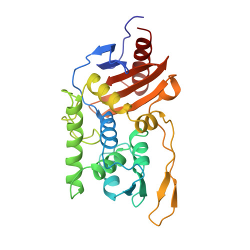
The serine DD-transpeptidase/penicillin-binding protein of Streptomyces K15 catalyzes peptide bond formation in a way that mimics the penicillin-sensitive peptide cross-linking reaction involved in bacterial cell wall peptidoglycan assembly. The Streptomyces K15 enzyme is peculiar in that it can be considered as an intermediate between classical penicillin-binding proteins, for which benzylpenicillin is a very efficient inactivator, and the resistant penicillin-binding proteins that have a low penicillin affinity. With its moderate penicillin sensitivity, the Streptomyces K15 DD-transpeptidase would be helpful in the understanding of the structure-activity relationship of this penicillin-recognizing protein superfamily. The structure of the Streptomyces K15 enzyme has been determined by x-ray crystallography at 2.0-A resolution and refined to an R-factor of 18.6%. The fold adopted by this 262-amino acid polypeptide generates a two-domain structure that is close to those of class A beta-lactamases. However, the Streptomyces K15 enzyme has two particular structural features. It lacks the amino-terminal alpha-helix found in the other penicilloyl-serine transferases, and it exhibits, at its surface, an additional four-stranded beta-sheet. These two characteristics might serve to anchor the enzyme in the plasma membrane. The overall topology of the catalytic pocket of the Streptomyces K15 enzyme is also comparable to that of the class A beta-lactamases, except that the Omega-loop, which bears the essential catalytic Glu(166) residue in the class A beta-lactamases, is entirely modified. This loop adopts a conformation similar to those found in the Streptomyces R61 DD-carboxypeptidase and class C beta-lactamases, with no equivalent acidic residue.
Organizational Affiliation:
Centre d'Ingénierie des Protéines, Université de Liège, Institut de Physique, B5, Sart Tilman B-4000, Belgium. Eveline.Fonze@ulg.ac.be