1SJ9

Crystal structure of the uridine phosphorylase from Salmonella typhimurium at 2.5A resolution


Experimental Data Snapshot

  • Method: X-RAY DIFFRACTION
  • Resolution: 2.50 Å
  • R-Value Free: 0.279 
  • R-Value Work: 0.221 
  • R-Value Observed: 0.221 

wwPDB Validation   3D Report Full Report


This is version 1.4 of the entry. See complete history


Literature

Preliminary investigation of the three-dimensional structure of Salmonella typhimurium uridine phosphorylase in the crystalline state.

Dontsova, M.V.Gabdoulkhakov, A.G.Molchan, O.K.Lashkov, A.A.Garber, M.B.Mironov, A.S.Zhukhlistova, N.E.Morgunova, E.Y.Voelter, W.Betzel, C.Zhang, Y.Ealick, S.E.Mikhailov, A.M.

(2005) Acta Crystallogr Sect F Struct Biol Cryst Commun 61: 337-340

  • DOI: https://doi.org/10.1107/S1744309105007463
  • Primary Citation of Related Structures:  
    1SJ9

  • PubMed Abstract: 

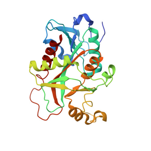
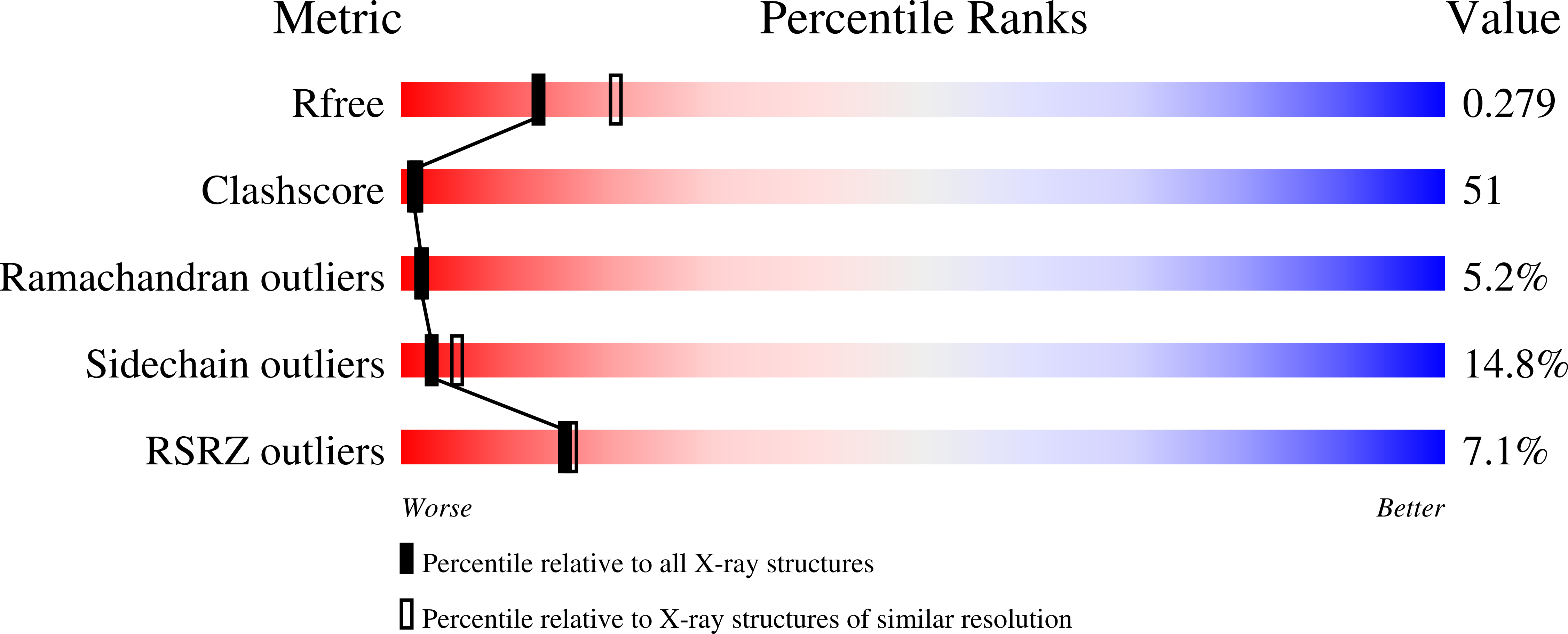
    Uridine phosphorylase (UPh) catalyzes the phosphorolytic cleavage of the C-N glycosidic bond of uridine to ribose 1-phosphate and uracil in the pyrimidine-salvage pathway. The crystal structure of the Salmonella typhimurium uridine phosphorylase (StUPh) has been determined at 2.5 A resolution and refined to an R factor of 22.1% and an Rfree of 27.9%. The hexameric StUPh displays 32 point-group symmetry and utilizes both twofold and threefold non-crystallographic axes. A phosphate is bound at the active site and forms hydrogen bonds to Arg91, Arg30, Thr94 and Gly26 of one monomer and Arg48 of an adjacent monomer. The hexameric StUPh model reveals a close structural relationship to Escherichia coli uridine phosphorylase (EcUPh).


  • Organizational Affiliation

    Institute of Protein Research, Russian Academy of Sciences, Pushchino, Russia.


Macromolecules
Find similar proteins by:  (by identity cutoff)  |  3D Structure
Entity ID: 1
MoleculeChains Sequence LengthOrganismDetailsImage
Uridine phosphorylase
A, B, C, D, E
A, B, C, D, E, F
253Salmonella enterica subsp. enterica serovar Typhimurium str. LT2Mutation(s): 0 
Gene Names: udp
EC: 2.4.2.3
UniProt
Find proteins for P0A1F6 (Salmonella typhimurium (strain LT2 / SGSC1412 / ATCC 700720))
Explore P0A1F6 
Go to UniProtKB:  P0A1F6
Entity Groups  
Sequence Clusters30% Identity50% Identity70% Identity90% Identity95% Identity100% Identity
UniProt GroupP0A1F6
Sequence Annotations
Expand
  • Reference Sequence
Experimental Data & Validation

Experimental Data

  • Method: X-RAY DIFFRACTION
  • Resolution: 2.50 Å
  • R-Value Free: 0.279 
  • R-Value Work: 0.221 
  • R-Value Observed: 0.221 
  • Space Group: P 61
Unit Cell:
Length ( Å )Angle ( ˚ )
a = 91.37α = 90
b = 91.37β = 90
c = 266.38γ = 120
Software Package:
Software NamePurpose
CNSrefinement
MAR345data collection
XDSdata scaling
MOLREPphasing

Structure Validation

View Full Validation Report


Entry History 

Deposition Data

Revision History  (Full details and data files)

  • Version 1.0: 2005-03-08
    Type: Initial release
  • Version 1.1: 2008-05-01
    Changes: Version format compliance
  • Version 1.2: 2011-07-13
    Changes: Derived calculations, Source and taxonomy, Version format compliance
  • Version 1.3: 2017-10-11
    Changes: Refinement description
  • Version 1.4: 2023-08-23
    Changes: Data collection, Database references, Derived calculations, Refinement description