Exploring sequence/folding space: folding studies on multiple hydrophobic core mutants of ubiquitin
Benitez-Cardoza, C.G., Stott, K., Hirshberg, M., Went, H.M., Woolfson, D.N., Jackson, S.E.(2004) Biochemistry 43: 5195-5203
- PubMed: 15122885
- DOI: https://doi.org/10.1021/bi0361620
- Primary Citation of Related Structures:
1SIF - PubMed Abstract:
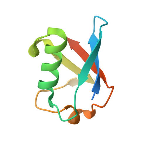
The stability, dynamic, and structural properties of ubiquitin and two multiple hydrophobic core mutants were studied. One of the mutants (U4) has seven substitutions in the hydrophobic core (M1L, I3L, V5I, I13F, L15V, V17M, and V26L). On average, its side chains are larger than the wild-type, and it can thus be thought of as having an overpacked core. The other mutant (U7) has two substitutions (I3V and I13V). On average, it has smaller side chains than the wild-type, and it can therefore be considered to be underpacked. The three proteins are well-folded and show similar backbone dynamics (T(1), T(2), and HNOE values), indicating that the regular secondary structure extends over the same residue ranges. The crystallographic structure of U4 was determined. The final R(factor) and R(free) are 0.198 and 0.248, respectively, at 2.18 A resolution. The structure of U4 is very similar to wild-type ubiquitin. Remarkably, there are almost no changes in the positions of the C(alpha) atoms along the entire backbone, and the hydrogen-bonding network is maintained. The mutations of the hydrophobic core are accommodated by small movements of side chains in the core of mutated and nonmutated residues. Unfolding and refolding kinetic studies revealed that U4 unfolds with the highest rates; however, its refolding rate constants are very similar to those of the wild-type protein. Conversely, U7 seems to be the most destabilized protein; its refolding rate constant is smaller than the other two proteins. This was confirmed by stopped-flow techniques and by H/D exchange methodologies. This work illustrates the possibility of repacking the hydrophobic core of small proteins and has important implications in the de novo design of stable proteins.
Organizational Affiliation:
University of Cambridge, Chemistry Department, Lensfield Road, Cambridge, CB2 1EW, United Kingdom.