1SHV

STRUCTURE OF SHV-1 BETA-LACTAMASE


Experimental Data Snapshot

  • Method: X-RAY DIFFRACTION
  • Resolution: 1.98 Å
  • R-Value Free: 0.254 
  • R-Value Work: 0.182 

wwPDB Validation   3D Report Full Report


This is version 1.4 of the entry. See complete history


Literature

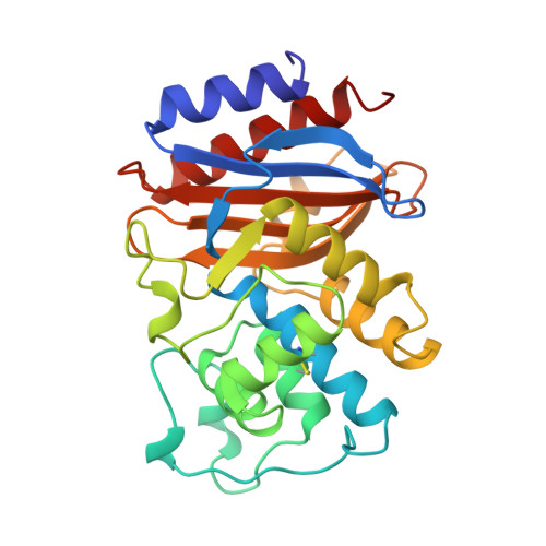
Structure of the SHV-1 beta-lactamase.

Kuzin, A.P.Nukaga, M.Nukaga, Y.Hujer, A.M.Bonomo, R.A.Knox, J.R.

(1999) Biochemistry 38: 5720-5727

  • DOI: https://doi.org/10.1021/bi990136d
  • Primary Citation of Related Structures:  
    1SHV

  • PubMed Abstract: 

    The X-ray crystallographic structure of the SHV-1 beta-lactamase has been established. The enzyme crystallizes from poly(ethylene glycol) at pH 7 in space group P212121 with cell dimensions a = 49.6 A, b = 55.6 A, and c = 87.0 A. The structure was solved by the molecular replacement method, and the model has been refined to an R-factor of 0.18 for all data in the range 8.0-1.98 A resolution. Deviations of model bonds and angles from ideal values are 0.018 A and 1.8 degrees, respectively. Overlay of all 263 alpha-carbon atoms in the SHV-1 and TEM-1 beta-lactamases results in an rms deviation of 1.4 A. Largest deviations occur in the H10 helix (residues 218-224) and in the loops between strands in the beta-sheet. All atoms in residues 70, 73, 130, 132, 166, and 234 in the catalytic site of SHV-1 deviate only 0.23 A (rms) from atoms in TEM-1. However, the width of the substrate binding cavity in SHV-1, as measured from the 104-105 and 130-132 loops on one side to the 235-238 beta-strand on the other side, is 0.7-1.2 A wider than in TEM-1. A structural analysis of the highly different affinity of SHV-1 and TEM-1 for the beta-lactamase inhibitory protein BLIP focuses on interactions involving Asp/Glu104.


  • Organizational Affiliation

    Department of Molecular and Cell Biology, The University of Connecticut, Storrs 06269-3125, USA.


Macromolecules
Find similar proteins by:  (by identity cutoff)  |  3D Structure
Entity ID: 1
MoleculeChains Sequence LengthOrganismDetailsImage
PROTEIN (BETA-LACTAMASE SHV-1)265Klebsiella pneumoniaeMutation(s): 0 
Gene Names: BLA
EC: 3.5.2.6
UniProt
Find proteins for P0AD64 (Klebsiella pneumoniae)
Explore P0AD64 
Go to UniProtKB:  P0AD64
Entity Groups  
Sequence Clusters30% Identity50% Identity70% Identity90% Identity95% Identity100% Identity
UniProt GroupP0AD64
Sequence Annotations
Expand
  • Reference Sequence
Small Molecules
Ligands 1 Unique
IDChains Name / Formula / InChI Key2D Diagram3D Interactions
MA4
Query on MA4

Download Ideal Coordinates CCD File 
B [auth A]CYCLOHEXYL-HEXYL-BETA-D-MALTOSIDE
C24 H44 O11
WUCWJXGMSXTDAV-QKMCSOCLSA-N
Experimental Data & Validation

Experimental Data

  • Method: X-RAY DIFFRACTION
  • Resolution: 1.98 Å
  • R-Value Free: 0.254 
  • R-Value Work: 0.182 
  • Space Group: P 21 21 21
Unit Cell:
Length ( Å )Angle ( ˚ )
a = 49.6α = 90
b = 55.6β = 90
c = 87γ = 90
Software Package:
Software NamePurpose
X-GENdata scaling
X-GENdata reduction
AMoREphasing
X-PLORmodel building
X-PLORrefinement
X-PLORphasing

Structure Validation

View Full Validation Report


Entry History 

Deposition Data

Revision History  (Full details and data files)

  • Version 1.0: 1999-05-06
    Type: Initial release
  • Version 1.1: 2007-10-16
    Changes: Version format compliance
  • Version 1.2: 2011-07-13
    Changes: Version format compliance
  • Version 1.3: 2017-10-04
    Changes: Refinement description
  • Version 1.4: 2023-08-23
    Changes: Data collection, Database references, Derived calculations, Refinement description