1SGV

STRUCTURE OF TRNA PSI55 PSEUDOURIDINE SYNTHASE (TRUB)


Experimental Data Snapshot

  • Method: X-RAY DIFFRACTION
  • Resolution: 1.90 Å
  • R-Value Free: 0.239 
  • R-Value Work: 0.201 
  • R-Value Observed: 0.203 

wwPDB Validation   3D Report Full Report


This is version 1.3 of the entry. See complete history


Literature

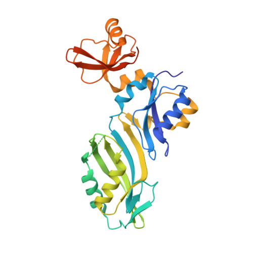
Crystal structure of the apo forms of psi 55 tRNA pseudouridine synthase from Mycobacterium tuberculosis: a hinge at the base of the catalytic cleft.

Chaudhuri, B.N.Chan, S.Perry, L.J.Yeates, T.O.

(2004) J Biol Chem 279: 24585-24591

  • DOI: https://doi.org/10.1074/jbc.M401045200
  • Primary Citation of Related Structures:  
    1SGV

  • PubMed Abstract: 

    The three-dimensional structure of the RNA-modifying enzyme, psi55 tRNA pseudouridine synthase from Mycobacterium tuberculosis, is reported. The 1.9-A resolution crystal structure reveals the enzyme, free of substrate, in two distinct conformations. The structure depicts an interesting mode of protein flexibility involving a hinged bending in the central beta-sheet of the catalytic module. Key parts of the active site cleft are also found to be disordered in the substrate-free form of the enzyme. The hinge bending appears to act as a clamp to position the substrate. Our structural data furthers the previously proposed mechanism of tRNA recognition. The present crystal structure emphasizes the significant role that protein dynamics must play in tRNA recognition, base flipping, and modification.


  • Organizational Affiliation

    UCLA-Department of Energy Institute for Genomics and Proteomics, Los Angeles, California 90095, USA.


Macromolecules
Find similar proteins by:  (by identity cutoff)  |  3D Structure
Entity ID: 1
MoleculeChains Sequence LengthOrganismDetailsImage
tRNA pseudouridine synthase B
A, B
316Mycobacterium tuberculosis H37RvMutation(s): 0 
Gene Names: TRUBRV2793CMT2862.1MTV002.58CMB2816C
EC: 4.2.1.70
UniProt
Find proteins for P9WHP7 (Mycobacterium tuberculosis (strain ATCC 25618 / H37Rv))
Explore P9WHP7 
Go to UniProtKB:  P9WHP7
Entity Groups  
Sequence Clusters30% Identity50% Identity70% Identity90% Identity95% Identity100% Identity
UniProt GroupP9WHP7
Sequence Annotations
Expand
  • Reference Sequence
Experimental Data & Validation

Experimental Data

  • Method: X-RAY DIFFRACTION
  • Resolution: 1.90 Å
  • R-Value Free: 0.239 
  • R-Value Work: 0.201 
  • R-Value Observed: 0.203 
  • Space Group: P 1 21 1
Unit Cell:
Length ( Å )Angle ( ˚ )
a = 45.948α = 90
b = 94.1β = 100.36
c = 78.508γ = 90
Software Package:
Software NamePurpose
REFMACrefinement
DENZOdata reduction
SCALEPACKdata scaling
AMoREphasing

Structure Validation

View Full Validation Report


Entry History 

Deposition Data

Revision History  (Full details and data files)

  • Version 1.0: 2004-03-02
    Type: Initial release
  • Version 1.1: 2008-04-29
    Changes: Version format compliance
  • Version 1.2: 2011-07-13
    Changes: Advisory, Source and taxonomy, Version format compliance
  • Version 1.3: 2023-08-23
    Changes: Data collection, Database references, Refinement description